Post-translational Modifications
реклама
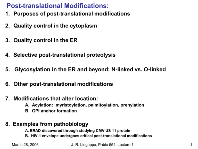
Post-translational Modifications: 1. Purposes of post-translational modifications 2. Quality control in the cytoplasm 3. Quality control in the ER 4. Selective post-translational proteolysis 5. Glycosylation in the ER and beyond: N-linked vs. O-linked 6. Other post-translational modifications 7. Modifications that alter location: A. Acylation: myristoylation, palmitoylation, prenylation B. GPI anchor formation 8. Examples from pathobiology A. ERAD discovered through studying CMV US 11 protein B. HIV-1 envelope undergoes critical post-translational modifications March 28, 2006 J. R. Lingappa, Pabio 552, Lecture 1 1 Post-translational Modifications: 1. Review of Translation: March 28, 2006 J. R. Lingappa, Pabio 552, Lecture 1 2 Post-translational Modifications: 1. Purposes of Post-translational Events & Modifications: A. Quality Control: Chaperones, Glycosylation B. Degradation of misfolded proteins: Ubiquitination, ERAD C. Proper protein function: Glycosylation, Phosphorylation, Ubiquitination D. Target protein to proper locations: Acylation, GPI anchors March 28, 2006 J. R. Lingappa, Pabio 552, Lecture 1 3 Post-translational Modifications: 2. Quality Control in the Cytoplasm: A. Anfinsen's dogma: All information needed for folding contained in the amino acid sequence: Leads to the concept of spontaneous protein folding. B. Problems with Anfinsen's dogma (and the notion of spontaneous folding): Features of cellular environments cause misfolding & aggregation. 1. Some proteins take a very long time to fold spontaneously. 2. Some protein domains are prone to misfolding and aggregation. March 28, 2006 J. R. Lingappa, Pabio 552, Lecture 1 4 Post-translational Modifications: 2. Quality Control in the Cytoplasm: B. Protein folding in vivo Problems with Anfinsen's dogma, cont. Folding in the cell differs from refolding of a denatured protein in vitro due to: final folded structure nascent chain Vectorial nature of protein synthesis in vivo. Exposure of hydrophobic regions during synthesis. Translation happens more slowly than folding, requiring a “delay” mechanism to allow translation to "catch up". PRODUCTIVE PATHWAY aggregation due to exposure of hydrophobic regions Highly crowded cytoplasm: 300 mg/ml prot. Folding in vitro is inefficient (20 - 30%); in the cell, efficiency close to 100%. Conditions of stress found in vivo exacerbate misfolding and aggregation. DEAD-END PATHWAY March 28, 2006 J. R. Lingappa, Pabio 552, Lecture 1 5 Post-translational Modifications: 2. Quality Control in the Cytoplasm: C. Molecular Chaperones: Proteins that mediate correct fate of other polypeptides but are not part of the final structure. Fate includes folding, assembly, interaction with other cellular components, transport, or degradation. A. History: Molecular chaperones initially identified as heat shock proteins, i.e. proteins upregulated by heat shock and other stresses. Heat shock causes protein denaturation with exposure and aggregation of interactive surfaces. Heat shock proteins inhibit aggregation by binding to exposed surfaces during times of stress but also during normal protein synthesis Thus, the stress response is simply an amplification of a normal function that is used by cells under non-stress conditions. March 28, 2006 J. R. Lingappa, Pabio 552, Lecture 1 6 Post-translational Modifications: D. Features of molecular chaperones: i. Hsp 70 family members: 70 kD protein monomers. Include DnaJ (bacteria); BiP (ER) Stabilize polypeptide surfaces in an unfolded state. Bind transiently to newly-synthesized proteins: paradoxically, efficient folding requires "antifolding". Bind permanently to misfolded protein. Have affinity for exposed hydrophobic peptides. Do NOT bind a specific sequence. Present in bacteria, eukaryotes & all compartments. Regulated by ATP hydrolysis. Undergo cycles of binding and release Act with cofactors (i.e. DnaJ, GrpE, Hip, Hop, Bag1). March 28, 2006 J. R. Lingappa, Pabio 552, Lecture 1 Hsp 70 Hsp 70 stabilizes the nascent chain 7 Post-translational Modifications: D. Features of molecular chaperones: ii. Chaperonins (GroEL, Hsp 60, TCP-1): Facilitate proper folding Bind and hydrolyze ATP Bind transiently to 10-15% proteins, but 2-3fold more w/stress 60 kD proteins that form oligomeric, stacked double rings Bring non-native substrate protein to central cavity folding where protected from aggregation with other non-native proteins Cycles of binding and release until the protein is properly folded GroEL (prokaryotic hsp 60) uses a cofactor, GroES. iii. Others: I.e. small heat shock proteins, hsp 90, etc. March 28, 2006 J. R. Lingappa, Pabio 552, Lecture 1 8 Post-translational Modifications: iv. Cytosolic chaperone co-ordination: Chaperones act in tandem. Stabilization by Hsp 70 plus cofactors) may be followed by use of Hsp 60 for proper folding. From Frydman, J. Annual Rev. of Biochemistry 70:603, 2001 March 28, 2006 J. R. Lingappa, Pabio 552, Lecture 1 9 Post-translational Modifications: 3. Quality control in the ER: A. Translation and translocation of proteins into the ER: Proteins that translocate into ER of mammalian cells include secretory proteins, TM proteins, or residents of a membranous compartment. These are targeted to the ER CO-TRANSLATIONALLY by an N-terminal signal sequence that generally gets cleaved during translocation across the ER membrane. The Signal Hypothesis March 28, 2006 SRP and SRP Receptor J. R. Lingappa, Pabio 552, Lecture 1 10 Post-translational Modifications: Translocation of Secretory Protein Translocation of Single Pass TM Protein March 28, 2006 Translocation of Double Pass TM Protein J. R. Lingappa, Pabio 552, Lecture 1 11 Post-translational Modifications: 3. Quality Control in the ER: B. Features of the ER: Proteins need to be unfolded to translocate Until signal sequence cleaved, N terminus of protein is constrained "incorrectly” ER lumen is topologically equivalent to extracellular space High oxidizing potential (unlike cytoplasm which is highly reduced) High Ca+2 concentration unlike cytoplasm Many sugars present along with machinery for glycosylation As in cytoplasm: high protein conc. (100 mg/ml) promotes aggregation As in cytoplasm: delay between translation/ translocation vs. folding Site of specific post-translational events: signal cleavage, S-S bond formation, N-linked glycosylation and GPI anchor addition March 28, 2006 J. R. Lingappa, Pabio 552, Lecture 1 12 Post-translational Modifications: 3. Quality Control in the ER: C. Specific ER chaperones: i. HSP 70 family members: BiP/GRP78 Recognize hydrophobic sequences in nascent chains. Undergo successive rounds of ATP-dependent binding and release. Essential for translocation of newly-synthesized proteins across the ER lumen and for retrograde transport into the cytosol (see ERAD, below). ii. Immunophilins/ FKBP - peptidyl prolyl isomerases. iii. Thiol-disulfide isomerases - PDI and ERp57 iv. Calnexin and Calreticulin: Unique to the ER Are lectins (carbohydrate binding proteins) Calreticulin - lumenal; Calnexin - integral membrane protein March 28, 2006 J. R. Lingappa, Pabio 552, Lecture 1 13 Post-translational Modifications: 3. Quality Control in the ER D. Mechanisms To pass QC checkpoints, protein must be correctly folded (most energetically favorable, native state) If protein fails to fold properly it must be degraded I. Example 1: BiP BiP (Hsp70 in ER) binds to newly-synthesized and unfolded chains. BiP stays associated with misfolded (but not properly folded) proteins. Retention by BiP leads to degradation (see proteolysis below). March 28, 2006 J. R. Lingappa, Pabio 552, Lecture 1 14 Post-translational Modifications: 3. Quality Control in the ER D. Mechanisms, cont. ii. Example 2: Calnexin/calreticulin bind to incompletely folded monoglucosylated glycans Cycles of binding/release controlled by: Glucosidase II: cleaves glucose from core glycan UDP-glucose: glucosyltransferase (GT) reglucosylates incompletelyfolded proteins so that they bind lectins again Thus GT acts as a folding sensor: proteins exit the cycle when GT fails to re-glucosylate. Glucose is a tag that signifies incomplete folding March 28, 2006 J. R. Lingappa, Pabio 552, Lecture 1 15 Post-translational Modifications: 3. Quality Control in the ER D. Mechanisms, cont. iii. Example 3: Trimming of a single mannose is a signal for degradation. Causes association with ER degradationenhancing mannosidase like protein (EDEM), which is a link to ER-associated degradation (see proteolysis below) Tsai, B. et al. Nature Rev. Mol. Cell Bio. 3: 246 (2002). March 28, 2006 J. R. Lingappa, Pabio 552, Lecture 1 16 Post-translational Modifications: 4. Selective post-translational proteolysis. Selective proteolysis is critical for cellular regulation. 3 steps for proteolysis in the cytoplasm: identify protein to be degraded mark it by ubiquitination deliver it to the proteasome, a protease complex that degrades it A. The Ubiquitin/Proteasome system: Ubiquitin: A highly-conserved 76 aa protein present in all eukaryotes. Covalently attached to e-amino groups in lysine side chains, Can be a single ubiquitin or multiple branched ubiquitins. Signal for ubiquitination can be: 1. Programmed via hydrophobic sequence or other motif 2. Acquired by 1) phosphorylation, 2) binding to adaptor protein, or 3) protein damage due to fragmentation, oxidation or aging. March 28, 2006 J. R. Lingappa, Pabio 552, Lecture 1 17 Post-translational Modifications: 4. Post-translational Quality Control: Selective proteolysis. B. Ubiquitination requires 3 enzymes: E1 (ubiquitin-activating enzyme) activates ubiquitin (U) E2 (ubiquitin-conjugating enzyme) acquires U via high-energy thioester E3 (ubiquitin ligase) transfers U to target proteins Hierarchical organization: one or few E1s exist, more E2s, many E3s. Other functions for ubiquitination (to be discussed in plasma membrane lecture). March 28, 2006 J. R. Lingappa, Pabio 552, Lecture 1 18 Post-translational Modifications: 4. Post-translational Quality Control: Selective proteolysis B. The Proteasome - high molecular weight (28S) protease complex that degrades ubiquitinated proteins in the cytoplasm Present in cytoplasm and nucleus, not ER Uses ATP Contains a 700 kD protease core and two 900 kD regulatory domains. Highly conserved and similar to proteases found in bacteria. Shaped like a cylinder. Proteins enter the cavity, and are cleaved into small peptides. Most but not all proteasome substrates are ubiqutinated. March 28, 2006 J. R. Lingappa, Pabio 552, Lecture 1 19 Post-translational Modifications: 4. Post-translational Quality Control: Selective Proteolysis C. Misfolding in the ER results in: ER-associated degradation (see below) Lysosomal degradation (next lecture) ER-Associated Protein Degradation (ERAD): Earlier notion was that ER had proteases. However, in fact most ER proteins targeted for degradation undergo retrograde translocation into cytosol and delivery to the proteasome. ER-Associated Degradation (ERAD) U U cytoplasm U cytoplasm ATP U U U U ER lumen misfolded protein hsp 70 (BiP) ER lumen U U translocon proteasome U ubiquitin March 28, 2006 J. R. Lingappa, Pabio 552, Lecture 1 20 Post-translational Modifications: 5. Glycosylation in the ER and beyond: Role of sugars in the ER: bulky hydrophilic groups that maintain proteins in solution, affect protein conformation, and allow lectins to facilitate folding and exert quality control. A. N-linked glycosylation - co-translational; recognizes Asn-x-Ser/Thr on nascent chain Catalyzed by oligosaccharyltransferases - integral membrane proteins with active site in the lumen. Transfers a preformed "high mannose" 14-residue sugar(Glc3Man9GlcNAc2) en bloc to asparagine residues on the acceptor nascent polypeptide chains. Highly conserved in mammals, plants, fungi. i. Donor molecule is dolichol-P-P-Glc3Man9GlcNAc2. Dolichol is a very long lipid carrier. ii. Subsequent trimming of residues (also called processing) off core sugar attached to protein occurs in the ER via glucosidases and mannosidases. N glycosylation can be prevented using: Tunicamycin: inhibits formation of the dolichol-P-P precursor. March 28, 2006 J. R. Lingappa, Pabio 552, Lecture 1 21 Post-translational Modifications: Bacteria: no N-glycosylation via dolichol 5. Glycosylation in the ER and Yeast: have only oligomannose type Nbeyond: glycans, because they don't have the ability A. N-linked glycosylation, cont. iii. -Glucosyltransferase recognizes misfolded glycoproteins and reglycosylates them. iv. Calreticulin and calnexin serve as sensors by binding to monoglucosylated proteins, facilitating their folding and assembly. v. Only glycoproteins that have been correctly folded (by calnexin and calreticulin), get packaged into ER-toGolgi transport vesicles. vi. In the cis Golgi, further processing & addition of GlcNac's to form branched structures vii. Addition of more sugar residues in the trans-Golgi (I.e. fucose and sialic acid) to produce the diversity that is seen in mature glycans. March 28, 2006 to add GlcNac in the trans Golgi Since bacteria & yeast lack Glc-Nac transferase enzyme, this enzyme demarcates a fundamental evolutionary boundary between uni- and multicellular organisms. J. R. Lingappa, Pabio 552, Lecture 1 22 Post-translational Modifications: Simplified view of N-glycosylation 4. monoglucosylated proteins are bound and folded by calnexin and calreticulin 1. core sugar added en bloc co-translationally to asparagine residues in nascent chains (from dolichol donor) 2. trimming of glucose residues in ER 3 glucosyl transferase adds back glucose in ER to unfolded glycoproteins = Glucose = Mannose = GlcNac = Galactose = Sialic Acid 6. in the medial and trans-Golgi more N-acetylglucosamines and fucose are added as well as galactoses and sialic acid (terminal glycosylation) using GlcNac transferase March 28, 2006 5. in the Golgi, trimming of mannose residues occurs medial-Golgi cis-Golgi J. R. Lingappa, Pabio 552, Lecture 1 23 Post-translational Modifications: 5. Glycosylation in the ER and beyond: B. O-linked glycosylation Many different types of sugars are added onto -OH of serine or threonine residues. Most of these are added in ER or Golgi However, addition of N-acetylglucosamine (GlcNac) can occur in cytoplasm on many different types of proteins May play an important role in signaling, much like phosphorylation May act in signaling to oppose phosphorylation March 28, 2006 J. R. Lingappa, Pabio 552, Lecture 1 24 Post-translational Modifications: 6. Other post-translational modifications: A. Disulfide bond formation in the ER Protein disulfide isomerase (PDI): in the ER: catalyzes oxidation of disulfide bonds in the cytosol and at the plasma membrane: reduces disulfide bonds Other proteins that act like PDI may be even more important in disulfide bond formation Requires action of a regenerating molecule (i.e. glutathione); NADPH is the source of redox equivalents. Disulfide Bond Formation SH S PDI substrate S SH S redox regenerator S S SH substrate SH redox regenerator PDI S SH SH March 28, 2006 J. R. Lingappa, Pabio 552, Lecture 1 25 Post-translational Modifications: 6. Other post-translational modifications, cont. B. Phosphorylation Kinases phosphorylate proteins at the hydroxyl groups of serine, threonine, and tyrosine Occurs in cytoplasm and nucleus C. Intracellular Proteolytic Cleavage Furin - protease that cleaves specific sites, located in the transGolgi network and in endosomes. D. Modified amino acids: hydroxyproline, hydroxylysine, 3-methylhistidine E. Lipidation March 28, 2006 J. R. Lingappa, Pabio 552, Lecture 1 26 Post-translational Modifications: 7. Post-translational Modifications that Alter Location: A. Acylation - Lipid attachments that anchor proteins to the membranes: Include myristoylation, palmitoylation, prenylation Involves addition to protein of fatty acids (long hydrocarbon ending in COOH) Allows proteins to target to the cytoplasmic faces of membrane compartments March 28, 2006 J. R. Lingappa, Pabio 552, Lecture 1 27 Post-translational Modifications: 7. Post-translational Modifications that Alter Location: i. Myristoylation: addition of C-14 FA myristate to N-terminus in cytoplasm Donor is myristoyl CoA Occurs co-translationally in the cytoplasm; can occur post-translationally when hidden motif is revealed by protein cleavage (i.e. pro-apoptotic protein BID) Enzyme NMT recognizes consensus sequence at N-terminus often revealed by a conformational change (myristoyl switch). Promotes weak but typically irreversible interaction with cytosolic membrane face Myristoylated proteins traffic through the cytoplasm Myristoylation necessary but not sufficient for membrane binding Second signal needed for membrane binding: myristate plus basic (basic aa’s interact with acidic phospholipids PS and PI), or myristate plus palmitate Met Gly Myristoylation Removal of initiating methionine Gly N-myristoyltransferase (NMT) Addition of myristate to N-terminal CH 3 March 28, 2006 O C-N-CH 2-C H Gly J. R. Lingappa, Pabio 552, Lecture 1 28 Post-translational Modifications: 7. Post-translational Modifications that Alter Location: ii. Palmitoylation - addition of a C-16 fatty acid to the thiol side chain of an internal cysteine residue. Promotes a reversible interaction with membrane Palmitoylated proteins traffic to membrane via cytoplasm or via secretory pathway Enzymes not well understood Myristoylated and palmitoylated proteins are enriched in caveolae and rafts Palmitoylation Cys SH2 Cys CH 3 S C H O March 28, 2006 J. R. Lingappa, Pabio 552, Lecture 1 29 Post-translational Modifications: 7. Post-translational Modifications that Alter Location: iii. Prenylation - addition of prenyl groups (two types) to S in internal cysteine a. Farnesylation - C15 fatty acid to C terminus by thioester linkage Occurs at CAAX sequences: cys, 2 aliphatic residues and C-terminal residue After attachment, last 3 residues are removed and new C terminal methylated Creates a highly hydrophobic C terminus b. Geranylgeranylation - similar to above but addition of C-20 to C terminal Cys Cys Farnesylation A A X A A X SH addition of farnesyl group Cys S proteolysis Cys S methylation Cys S March 28, 2006 J. R. Lingappa, Pabio 552, Lecture 1 -O-CH 3 30 Post-translational Modifications: 7. Post-translational Modifications that Alter Location: iii. Examples of acylated proteins important for pathogenesis: Myristoylated proteins: HIV-1 Gag, HIV-1 Nef which target to the PM; Arfs involved in coat protein binding to vesicles (see ER-Golgi lecture) Palmitoylated proteins: caveolin (see PM lecture) Dual acylated proteins (myr plus palm): found in Src tyrosine kinases, i.e. Lyn, Fyn, Hck, etc. (see Signaling overview lecture) Met-Gly-Cys signal for dual acylation Farnesylation: Ras, does not insert into the membrane or act in signal transduction unless farnesylated. Geranylgeranylation: Rab GTP-binding proteins that mediate initial vesicle targeting events (see PM lecture) March 28, 2006 J. R. Lingappa, Pabio 552, Lecture 1 31 Post-translational Modifications: 7. Post-translational Modifications that Alter Location: B. GPI anchors - Glycophosphatidyl inositol (GPI) attached to the C terminus Composed of oligosaccharides and inositol phospholipids Provides a mechanism for anchoring cell-surface proteins to the membrane as a flexible leash that allows the entire protein (except for anchor) to be in extracellular space (unlike a transmembrane protein) Added to translocated proteins in ER Targets to PM via secretory pathway Unlike with N- or O-glycosylation, no more than ONE GPI anchor per protein Unlike acylation, targets proteins to outer leaflet of plasma membrane Can be cleaved by PI-phospholipase C (PI-PLC) Are minor components on mammalian cells but abundant on surfaces of parasitic protozoa (i.e. trypanosomes and Leishmania) and yeasts Concentrated in lipid rafts March 28, 2006 J. R. Lingappa, Pabio 552, Lecture 1 32 Post-translational Modifications: Structure of a GPI anchor: Protein C=O NH CH2 CH2 N-Acetylgalactosamine C-terminus ETHANOLAMINE P Mannose OLIGOSACCHARIDE NH3 CH2 Lipid Bilayer March 28, 2006 CH2 P Glucosamine Inositol head of PHOSPHATIDYLINOSITOL J. R. Lingappa, Pabio 552, Lecture 1 33 Post-translational Modifications: 7. Post-translational Modifications that Alter Location: B. GPI anchors - Functions: Stronger anchoring to PM than acylation Some GPI anchors can be replaced with TM anchors and be functional; others cannot Crosslinking results in signal transdcution across bilayer, including Ca influx, tyrosine phosphorylation, cytokine secretion, etc. Can interact with TM proteins capable of intracellular signaling Can indirectly modulate activity of cytosolic signaling molecules assoc. w/ lipid rafts March 28, 2006 GPI Anchor Formation GPI anchored protein tethered to outer leaflet of PM cytoplasm ER lumen protein translation and translocation ER cytoplasm extracellular space GPI cytoplasm GPI cleavage of hydrophobic C terminal sequence and transfer of preformed GPI moiety ER lumen ER cytoplasm PM extracellular space GPI PM cytoplasm ER lumen vesicle fusion ER GPI vesicle formation cytoplasm =N terminal signal sequence =C terminal GPI signal ER lumen vesicle transport ER J. R. Lingappa, Pabio 552, Lecture 1 34 Post-translational Modifications: 8. Examples from Pathobiology: A. ERAD discovered through study of CMV US11 (Wiertz et al., Cell 84: 769, 1996). 1. MHC class I, a TM protein, binds viral peptides produced in cells and presents them at the cell surface to cytotoxic T cells. 2. CMV evades the immune system by targeting MHC class I for destruction soon after it is synthesized and translocated into the ER. How does it do this? 3. CMV US11 protein expressed alone causes MHC class I destruction. 4. US 11 effect is sensitive to proteasome inhibitors and involves MHC class I localization to cytoplasm, implying movemnt of US 11 out of ER into cytoplasm for degradation. 5. Before this paper, only forward movement thru translocon was thought to occur; this paper by Ploegh’s group studying a CMV protein raised the possibility of retrograde movement thru translocon. ERAD: 6. Subsequently, retrograde movement thru translocon for degradation (ERAD) was shown to be a common in non-infected cells. 7. Note that MHC class I needs to be poly-ubiquitinated for retrograde transport to occur, implying a role for ubiqutination in retrolocation, not just in targeting for degradation. 8. Additional studies reveal that other pathogens use this mechanism: I.e. HIV-1 accessory protein Vpu promotes degradation of CD4 by ERAD. March 28, 2006 J. R. Lingappa, Pabio 552, Lecture 1 35 Post-translational Modifications: 8. Examples from Pathobiology: B. HIV-1 envelope protein undergoes many critical post-translational modifications 1. HIV env consists of gp120 soluble portion bound non-covalently to TM gp41. Role is to bind CD4 and chemokine receptors during HIV-1 entry. 2. Co-translationally translocated into ER as gp160. 3. Has ~30 potential sites for N-linked glycosylation in ER. If non-glycosylated: won’t bind CD4. Some glycosylations are dispensible for proper folding; others are needed. 4. Forms 10 disulfide bonds in ER (9 are in gp120 portion). 5. Trimerization of HIV-1 env in ER 6. Proper folding/trimerization equires BiP, calnexin, calreticulin, and PDI. 7. In Golgi: protease-mediated cleavage of gp160 to gp120 and gp41. March 28, 2006 Land, A. and I. Braakman, Biochimie 83: 783 (2001). J. R. Lingappa, Pabio 552, Lecture 1 36 Post-translational Modifications: Additional Reading: *Tsai, B. et al. Retro-translocation of proteins from the endoplasmic reticulum into the cytosol. Nature Rev. Mol. Cell Bio. 3: 246 (2002). Freiman, R. N. and R. Tijan. Regulating the regulators: Lysine modifications make their mark. Cell 112: 11 - 17 (2003). Resh, M. Fatty acylation of proteins: new insights into membrane targeting of myristoylated and palmitoylated proteins. BBA 1451: 1 (1999). Land, A. and I. Braakman. Folding of the human immunodeficiency virus type I envelope glycoprotein in the endoplasmic reticulum. Biochimie 83: 783 (2001). Chatterjee, S. and S. Mayor. The GPI-anchor and protein sorting. Cell Mol. Life Sci 58: 1969 (2001). McClellan A et al. Protein quality control: chaperones culling corrupt conformations. Nat Cell Biol. 2005 Aug;7(8):736-41. Gill, G. SUMO and ubiquitin in the nucleus: different functions, similar mechanisms? Genes Dev. 2004 Sep 1;18(17):2046-59. Review. March 28, 2006 J. R. Lingappa, Pabio 552, Lecture 1 37
