Dyes and Pigments 74 (2007) 665e671
www.elsevier.com/locate/dyepig
Spectroscopic investigations on the mechanism of interaction
of bioactive dye with bovine serum albumin
S.M.T. Shaikh, J. Seetharamappa*, P.B. Kandagal, D.H. Manjunatha, S. Ashoka
Department of Chemistry, Karnatak University, Dharwad 580 003, India
Received 7 January 2006; received in revised form 11 February 2006; accepted 24 April 2006
Available online 8 June 2006
Abstract
The mechanism of binding of rose bengal (RB) with bovine serum albumin (BSA) was investigated by spectroscopic methods. The analysis
of fluorescence data indicated the presence of both dynamic and static quenching mechanisms in the binding. Various binding parameters have
been evaluated. The thermodynamic parameters, DH0 and DS0 were observed to be 79.61 kJ mol1 and 143.37 J mol1 K1, respectively.
The quantitative analysis of CD results revealed that the a-helicity of BSA decreased from 66.4% (in free BSA) to 48.64% (in bound BSA).
The binding average distance, r between the BSA (donor) and RB (acceptor) was determined based on Förster’s theory and it was found to
be 2.75 nm. The effects of common ions on the binding constant of RBeBSA were also examined.
Ó 2006 Elsevier Ltd. All rights reserved.
Keywords: Bovine serum albumin; Rose bengal; Spectroscopic studies
1. Introduction
Dyes are being increasingly used for clinical and medicinal
purposes [1]. The discovery that some dyes would stain certain
tissues and not others led to the idea that dyes might destroy
pathogenic organisms without causing appreciable harm to
the host. Due to this, some azo, thiazine, triphenyl methane
and acridine dyes came into use as antiseptic trypanocides
and for other medicinal purposes [2]. RB, chemically,
4,5,6,7-tetrachloro 20 ,40 ,50 ,70 -tetraiodofluorescein di sodium
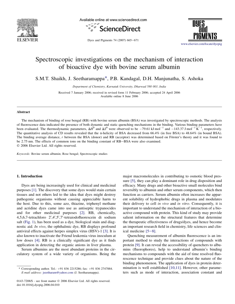
salt (Fig. 1), has been used as a dye, biological stain and diagnostic aid. In vivo, the ophthalmic dye, RB displays profound
antiviral effects against herpes simplex virus (HSV)-1 [3]. It is
also known to inactivate the Friend leukemia virus inoculum at
low doses [4]. RB is a clinically significant dye as it finds
application in detecting the organic anions in liver plasma.
Serum albumins are the most abundant proteins in the circulatory system of a wide variety of organisms. Being the
* Corresponding author. Tel.: þ91 836 2215286; fax: þ91 836 2747884.
E-mail address: jseetharam@yahoo.com (J. Seetharamappa).
0143-7208/$ - see front matter Ó 2006 Elsevier Ltd. All rights reserved.
doi:10.1016/j.dyepig.2006.04.010
major macromolecules in contributing to osmotic blood pressure [5], they can play a dominant role in drug disposition and
efficacy. Many drugs and other bioactive small molecules bind
reversibly to albumin and other serum components, which then
function as carriers. Serum albumin often increases the apparent solubility of hydrophobic drugs in plasma and modulates
their delivery to cell in vivo and in vitro. Consequently, it is
important to understand the mechanism of interaction of a bioactive compound with protein. This kind of study may provide
salient information on the structural features that determine
the therapeutic effectiveness of drugs/dyes, and hence become
an important research field in chemistry, life sciences and clinical medicine [5e8].
Quenching measurement of albumin fluorescence is an important method to study the interactions of compounds with
protein [9]. It can reveal the accessibility of quenchers to albumins (fluorophores), help to understand albumin’s binding
mechanisms to compounds with the aid of time resolved fluorescence technique and provide clues about the nature of the
binding phenomenon. The application of dyes in protein determination is well established [10,11]. However, other parameters such as mode of interaction, association constant and
S.M.T. Shaikh et al. / Dyes and Pigments 74 (2007) 665e671
666
I
I
O-Na+
O
I
O
Cl
O
3
2
1
Cl
Cl
0
Cl
0.00002
0.00001
0
[Q] M
Fig. 1. Structure of rose bengal.
Fig. 3. SterneVolmer plot for the binding of RB to BSA.
number of binding sites are important, when dyes are used as
drugs. The binding of dyes to proteins has seldom been investigated [12,13]. The literature survey reveals that attempts
have not been made so far to investigate the binding mechanism of RB with BSA by fluorescence, UVevis absorption,
circular dichroism and lifetime measurements. The energy
transfer between RB and protein (BSA) is also reported in
the present study for the first time.
2. Results and discussion
2.1. Fluorescence measurements
The fluorescence intensity of a compound can be decreased
by a variety of molecular interactions viz., excited-state reactions, molecular rearrangements, energy transfer, ground state
complex formation and collisional quenching. Such decrease
in intensity is called quenching. In order to investigate the
binding of RB to BSA, the fluorescence spectra were recorded
in the range of 300e500 nm upon excitation at 296 nm. RB
causes a concentration dependent quenching of the intrinsic
fluorescence of BSA (Fig. 2) without changing the emission
maximum and shape of the peaks. These results indicated
that there were interactions between RB and BSA. The interaction of RB with BSA was further confirmed by absorption
and circular dichroism techniques. The fluorescence quenching data are analyzed by the SterneVolmer equation,
F0 =F ¼ 1 þ KSV ½Q
ð1Þ
where F0 and F are the steady state fluorescence intensities in
the absence and presence of quencher, respectively, KSV is the
SterneVolmer quenching constant and [Q] is the concentration
of quencher (RB). The plot of F0/F versus [Q] showed positive
deviation (concave towards the Y axis) indicating the presence
of both static and dynamic quenching [14] by the same fluorophore (Fig. 3).
Fluorescence lifetimes were estimated for BSA from the
fluorescence decays (Fig. 4). A biexponential decay was observed with two nanosecond lifetimes, 6.08 and 2.08 ns [15].
Hence, due to the presence of two tryptophan residues of the
protein, the fluorescence decays are characterized by two
nanosecond lifetimes. Interestingly, the effect of addition of
drug to protein showed the quenching of both the lifetimes
as a function of the increase in the drug concentration. The
lifetimes (t1 and t2), relative amplitudes (A1 and A2) and c2
1.8
10000
b
To/T2
I
4
Fo/F
Na+O-
5
1.4
1
0.6
Log (counts)
120
Intensity
a
80
1000
0
100
200
300
400
[Q]
f
100
a
k
40
10
0
300
350
400
450
500
0
5
10
15
20
25
Time (ns)
Wavelength (nm)
Fig. 2. Fluorescence spectra of BSA in the presence of RB. BSA concentration
was fixed at 12 mM (a). RB concentrations were 2 (b), 4 (c), 6 (d), 8 (e), 10 (f),
12 (g), 14 (h), 16 (i), 18 (j), and 20 mM (k).
Fig. 4. Fluorescence decay profiles of BSA in the absence and presence of RB
in 0.1 M phosphate buffer of pH 7.4, lex ¼ 296 nm and lem ¼ 344 nm, (a) Laser profile, BSA concentration was fixed at 30 mM (b). In RBeBSA, RB concentrations were 70(c), 150 (d), 250 (e) and 300 mM (f).
S.M.T. Shaikh et al. / Dyes and Pigments 74 (2007) 665e671
7
of the various decay analysis of the BSAeRB system are listed
in Table 1. The dynamic portion of observed quenching was
determined by lifetime measurements using the equation,
ð2Þ
where t0 (6.08 ns) and t (equal to t2 in the present study) are
the fluorescence lifetimes of BSA in the absence and presence
of RB, respectively, and KD is the dynamic quenching constant. The value of KD was found to be 2.12 0.021 103 M1 from the plot of t0/t2 versus [Q]. The value of static
quenching constant, KS was obtained using the equation,
6
5
Fo/(Fo-F)
t0 =t ¼ 1 þ KD ½Q
4
3
2
1
0
F0 =ðF0 FÞ ¼ f1=fa þ 1g=ð½Qfa KSV Þ
ð4Þ
where fa is the fraction of the initial fluorescence which is accessible to the quencher and KSV is the SterneVolmer quenching constant. The value of fa refers to the fraction of
fluorescence accessible to quenching, which need not be the
same as the fraction of tryptophan residues i.e. accessible to
quenching [14]. From the plot of F0/(F0 F ) versus 1/[Q],
the values of fa and KSV were obtained from the values of intercept and slope, respectively (Fig. 5). The value of fa was observed to be 1.18 0.007 at 298 K indicating that 84 0.39%
of the total fluorescence of BSA is accessible to the quencher.
The KSV was found to be 7.43 0.051 104 M1.
2.2. Analysis of binding equilibria
When small molecules bind independently to a set of equivalent sites on a macromolecule, the equilibrium between free
and bound molecules is given by the equation [16],
log ðF0 FÞ=F ¼ log K þ n log½Q
ð5Þ
e
70
150
250
330
Biexp
Biexp
Biexp
Biexp
Biexp
0.3
0.4
0.5
log [Q] yielded the K and n values of 28.05 0.027 105 M1 and 1.1 0.016, respectively. The value of n is
approximately equal to 1 indicating that there is one class of
binding site to RB in BSA.
2.3. Type of interaction force between RB and BSA
The binding studies were carried out at 288, 298, 303 and
308 K. At these temperatures the BSA does not undergo any
structural degradation. The interaction between a ligand and
protein may involve hydrogen bonds, van der Waals forces,
electrostatic forces, and/or hydrophobic interactions. The thermodynamic parameters were determined using the following
equations,
log K ¼ DH0 =2:303RT þ DS0 =2:303R
ð6Þ
DG0 ¼ DH 0 TDS0
ð7Þ
The log K versus 1/T plot (Fig. 6) enabled the determination of DH0 and DS0 for the binding process. DH0, DG0 and
DS0 are standard enthalpy change, standard free energy change
and standard entropy change, respectively. The values of K,
DH0, DS0 and DG0 are summarized in Table 2. The negative
value of DG0 reveals that the interaction process is spontaneous. An important source of negative contribution to DH0 and
DS0 will arise if a hydrogen bond is formed [17]. The negative
DH0 and DS0 values for the interaction of RB and BSA indicate that the binding is mainly enthalpy driven and entropy
Lifetime (ns)
Amplitude
t1
t2
A1
A2
2.08
1.25
1.02
7.56
5.94
6.08
5.66
5.33
4.68
3.85
6.98
14.40
25.42
36.77
61.65
93.02
85.60
74.58
63.23
38.35
c2
7.2
6.8
Log K
Table 1
Lifetimes of fluorescence decay of BSA in phosphate buffer of pH 7.4 at different concentrations of RB
BSA (30 mM)
1
2
3
4
0.2
1/[Q]
where K and n are the binding constants and the number of
binding sites, respectively. Thus, a plot of log (F0 F )/F versus
Analysis
0.1
ð3Þ
by plotting the graph of {(F0 F )/F}/[Q] versus [Q] and
using the value of KD obtained from lifetime measurements.
It was found to be 4.76 0.023 104 M1. The fluorescence
data obtained at room temperature were further examined
using the modified SterneVolmer equation,
RB
(mM)
0
Fig. 5. Modified SterneVolmer plot for the binding of RB to BSA.
ðF0 FÞ=F
¼ ðKS þ KD Þ þ KS KD ½Q
½Q
Number
667
6.4
6
1.18
1.13
1.22
1.39
1.46
5.6
0.0032
0.0033
0.0034
1/T
Fig. 6. Log K versus 1/T for RBeBSA system.
0.0035
S.M.T. Shaikh et al. / Dyes and Pigments 74 (2007) 665e671
668
Table 2
Thermodynamic parameters of BSAeRB system
Drug
Temperature (K)
Binding constant (K 105, M1)
r
DG0 (kJ mol1)
DH0 (kJ mol1)
DS0 (J mol1 K1)
RB
288
298
303
308
(92.17 0.031)
(28.05 0.027)
(16.20 0.036)
(10.99 0.038)
0.9914
0.9982
0.9922
0.9896
38.38
37.40
35.96
35.61
79.61
143.37
is unfavorable for it, and that the hydrogen bonding and van
der Waals forces played major role in the interaction [17].
2.4. Energy transfer between RB and BSA
The overlap of the UV absorption spectra of RB with the
fluorescence emission spectra of BSA is shown in Fig. 7.
The energy transfer has been used as a ‘‘spectroscopic ruler’’
for the measurement of distance between the sites on proteins
[14]. The importance of energy transfer in biochemistry is
that, the efficiency of transfer can be used to evaluate the distance between the ligand and the tryptophan residues in the
protein. According to Förster’s non-radiative energy transfer
theory [18], the rate of energy transfer depends on: (i) the relative orientation of the donor and acceptor dipoles, (ii) the extent of overlap of fluorescence emission spectrum of the donor
with the absorption spectrum of the acceptor and (iii) the distance between the donor and the acceptor. The energy transfer
effect is related not only to the distance between the acceptor
and donor, but also to the critical energy transfer distance, R0,
that is
F
R6
E¼1 ¼ 6 0 6
F0 R0 þ r
ð8Þ
where F and F0 are the fluorescence intensities of BSA in the
presence and absence of RB, r is the distance between acceptor and donor and R0 is the critical distance when the transfer
efficiency is 50%. R60 is calculated using the equation,
R60 ¼ 8:8 1025 k2 N 4 FJ
ð9Þ
0.4
120
0.3
60
0.2
30
0
300
b
350
Absorbance
Intensity
a
90
0.1
400
450
0
500
Wavelength (nm)
Fig. 7. The overlap of the fluorescence spectrum of BSA (a) and the absorbance spectrum of RB (b) {lex ¼ 296 nm, lem ¼ 344 nm, [BSA]:[RB] ¼ 1:1}.
where k2 is the spatial orientation factor of the dipole, N is the
refractive index of the medium, F is the fluorescence quantum
yield of the donor and J is the overlap integral of the fluorescence emission spectrum of the donor and the absorption spectrum of the acceptor. J is given by
P
J¼
FðlÞ3ðlÞl4 Dl
P
FðlÞDl
ð10Þ
where F(l) is the fluorescence intensity of fluorescent donor of
wavelength, l, and 3(l) is the molar absorption coefficient of
the acceptor at wavelength, l. In the present case, k2 ¼ 2/3,
N ¼ 1.336 and F ¼ 0.15 [19,20]. From Eqs. (8)e(10), we
were able to calculate that J ¼ 1.52 1014 cm3 L M1,
R0 ¼ 1.86 nm, E ¼ 0.45 and r ¼ 2.75 nm. The donor-to-acceptor distance, r < 7 nm [21,22] indicated that the energy transfer from BSA to RB occurs with high possibility. Larger
BSAeRB distance, r compared to that of R0 value observed
in the present study also reveals the presence of static type
quenching mechanism to a larger extent [23,24].
2.5. The effect of ions on the binding constant of
RBeBSA
Since the common ions which are distributed in human and
animals (in ionized or non-ionized or complex form) do influence the binding of drugs to proteins, such studies have been
reported in the literature. Hence, we have examined the effects
of common ions viz., Fe2þ, Co2þ, Kþ, Ni2þ, V5þ and Cu2þ on
the binding constant of RBeBSA at 298 K. The solutions of
cations were prepared from chlorides of respective cations
while that of V5þ solution was prepared from ammonium
metavanadate. Under experimental conditions, no cation
gave precipitate in phosphate buffer. The binding constant
for RBeBSA increased in the presence of ions viz., Co2þ,
Kþ, Ni2þ and Cu2þ (Table 3) implying stronger binding of
Table 3
Effects of common ions on binding constants of BSAeRB system
System
Binding constant (M1)
BSA þ RB
BSA þ RB þ Fe2þ
BSA þ RB þ Co2þ
BSA þ RB þ Kþ
BSA þ RB þ V5þ
BSA þ RB þ Ni2þ
BSA þ RB þ Cu2þ
(28.05 0.027) 105
(14.21 0.041) 105
(32.74 0.038) 105
(81.11 0.047) 105
(10.13 0.026) 105
(87.09 0.019) 105
(84.02 0.035) 105
S.M.T. Shaikh et al. / Dyes and Pigments 74 (2007) 665e671
2.6. Conformation investigations
Observed CDðmdegÞ
Cp nl 10
ð11Þ
where CP is the molar concentration of the protein, n is the
number of amino acid residues and l is the path length. The
2
Absorbance
1.6
h
1.2
a
0.8
x
280
-5000
e
-10000
-15000
a
300
-25000
200
210
220
230
240
250
Wavelength (nm)
Fig. 9. CD spectra of the BSAeRB system obtained in 0.1 M phosphate buffer
of pH 7.4 at room temperature. BSA concentration was kept fixed at 12 mM
(a). In RBeBSA system, RB concentrations were 6 (b), 18 (c), 42 (d) and
90 mM (e).
a-helical contents of free and bound BSA [5] were calculated
from MRE value at 208 nm using the equation,
a-Helixð%Þ ¼
MRE208 4000
100
33; 000 4000
ð12Þ
where MRE208 is the observed MRE value at 208 nm, 4000 is
the MRE of the b-form and random coil conformation cross at
208 nm, and 33,000 is the MRE value of a pure a-helix at
208 nm. From the above equation, the quantitative analysis results of the a-helix in the secondary structure of BSA were obtained. They a-helicity differed from that of 66.41% in free
BSA to 48.64% in the BSAeRB system, which was indicative
of the loss of a-helical. The percentage of a-helical structure
of protein decreased indicating that RB bound with the amino
acid residues of the main polypeptide chain of protein and destroyed their hydrogen bonding networks [24]. The CD spectra
of BSA in presence and absence of RB are similar in shape,
indicating that the structure of BSA after RB binding to BSA
is also predominantly a-helical [27]. So, we concluded that
the binding of RB to BSA induced some conformational
changes in BSA.
3. Conclusions
0.4
0
260
0
-20000
UVevis absorption measurement is a very simple method
and applicable to explore the structural change [22] and to
know the complex formation [9]. In the present study, we
have recorded the UV absorption spectra of RB, BSA and
RBeBSA system (Fig. 8). It is evident that the UV absorption
intensity of BSA increased regularly with the variation of RB
concentration. The maximum peak position of RBeBSA was
shifted slightly towards lower wavelength region possibly
due to complex formation between RB and BSA [24]. The
slight shift of the peaks also indicated that with addition of
RB, the peptide strands of BSA molecules extended more
and the hydrophobicity was decreased [22].
CD is a sensitive technique to monitor the conformational
changes in the protein upon interaction with the ligand. The
CD spectra of BSA in the absence and presence of RB are
shown in Fig. 9. The CD spectra of BSA exhibited two negative bands in the UV region at 209 and 222 nm, characteristic
of an a-helical structure of protein [26]. The CD results were
expressed in terms of mean residue ellipticity (MRE) in
deg cm2 dmol1 according to the following equation [8],
MRE ¼
5000
[ ] (deg.cm2.dmol-1)
RB to BSA. This prolongs the storage time of RB in blood
plasma (if RB is administered to a subject in presence of
Co2þ, Kþ, Ni2þ and Cu2þ) and enhances the maximum effectiveness of the dye [24]. Hence, in the presence of above ions,
RB can be stored and removed better by BSA [24]. The decrease in binding constant of RBeBSA in the presence of
Fe2þ and V5þ (Table 3) revealed the availability of higher concentration of free RB in plasma which could be quickly
cleared from the blood through urine [25].
669
320
Wavelength (nm)
Fig. 8. Absorbance spectra of RB, BSA and BSAeRB system. BSA concentration was kept fixed at 12 mM (a). RB concentrations in RBeBSA system
were 7 (b), 14 (c), 21 (d) 28 (e), 35 (f), 49 (g) and 63 mM (h). A concentration
of 12 mM was used for RB only (x).
This paper provided an approach for studying the binding
of protein to RB by employing absorption, fluorescence, circular dichroism and lifetime measurements. The results showed
that the BSA fluorescence was quenched by RB through both
dynamic and static quenching. The spectral data revealed the
conformational changes in BSA upon interaction with RB.
The binding constant of RBeBSA was affected in presence
of common ions. This work also reports the distance between
670
S.M.T. Shaikh et al. / Dyes and Pigments 74 (2007) 665e671
the donor (BSA) and the acceptor (RB) for the first time based
on Förster’s energy transfer theory.
4. Experimental
4.1. Materials
Bovine serum albumin (BSA, Fraction V) was obtained
from Sigma Chemical Company, St Louis, USA. AnalaR
grade rose bengal was used in the study. The solutions of
RB and BSA were prepared in 0.1 M phosphate buffer of pH
7.4 containing 0.15 M NaCl. BSA solution was prepared based
on its molecular weight of 65,000. All other materials were of
analytical reagent grade and double distilled water was used
throughout.
4.2. Spectral measurements
Fluorescence measurements were performed on a Hitachi
spectrofluorimeter Model F-2000 equipped with a 150 W Xenon lamp and slit width of 5 nm. The CD measurements were
made on a JASCO-J-715 spectropolarimeter using a 0.1 cm
cell at 0.2 nm intervals, with three scans averaged for each
CD spectrum in the range of 200e250 nm. The absorption
spectra were recorded on a double beam CARY 50-BIO
UVevis spectrophotometer equipped with a 150 W Xenon
lamp and a slit width of 5 nm.
Fluorescence decays were recorded using standard timecorrelated single photon counting (TCSPC) method using
the following set up: a diode pumped millena continuous
wave (CW) laser (Spectra Physics) 532 nm was used to
pump the titanium:sapphire rod in Tsunami picoseconds
mode locked laser system (Spectra Physics). The 750 nm
(80 MHz) was taken from the titanium:sapphire laser and
passed through pulse picker (Spectra Physics, 3980 2S) to
generate 4 MHz pulses. The third harmonic output (296 nm)
was generated by flexible harmonic generator (Spectra Physics, GWU 23 PS). The vertically polarized 296 nm laser was
used to excite the sample. The fluorescence emission at
magic angle (54.7 ) was dispersed in a monochromator
( f/3 aperture), counted by a microchannel plate (MCP) photo
multiplier tube (PMT) [Hamamatsu R 3809] and processed
through CFD, TAC and microchannel analyzer (MCA). The
instrument response function for this system is w52 ps. Fluorescence decay was analyzed by using the software provided
by IBH (Decay Associated emission Spectra-6) and PTI
Global Analysis Software. A 1.0 cm quartz cell was used
throughout the study.
4.3. RBeBSA interactions
Based on preliminary experiments, BSA concentration was
kept fixed at 12 mM and drug concentration was varied from 2
to 20 mM. Fluorescence spectra were recorded at 288, 298, 303
and 308 K in the range of 300e500 nm upon excitation at
296 nm in each case.
4.4. Circular dichroism measurements
The CD measurements of BSA in the absence and presence
of RB (1:0.5, 1:1.5, 1:3.5 and 1:7.5) were made in the range of
200e250 nm. A stock solution of 150 mM BSA was prepared.
4.5. Lifetime measurements
The fluorescence lifetime measurements of BSA in the
presence and absence of RB were recorded by fixing 296 nm
as the excitation wavelength and 344 nm as the emission
wavelength. A stock solution of 200 mM BSA was prepared
in 0.1 M phosphate buffer of pH 7.4 containing 0.15 M
NaCl. The BSA concentration was kept fixed at 30 mM and
RB concentration was varied from 10 to 30 mM.
4.6. Effects of some common ions
The fluorescence spectra of RB (2e20 mM)eBSA (12 mM)
were recorded in the absence and presence of various common
ions (12 mM) viz., Fe2þ, Co2þ, Kþ, Ni2þ, V5þ and Cu2þ in the
range of 300e500 nm upon excitation at 296 nm.
Acknowledgements
The authors acknowledge Department of Science and Technology, New Delhi, India for financial support for this work
(SP/S1/H-38/2001). We thank Prof. M.R.N Murthy, Indian
Institute of Science, Bangalore, for CD measurement facilities.
We are also grateful to Prof. Ramamurthy, National Centre
for Ultrafast Processes, University of Madras, Chennai for
the lifetime measurements.
References
[1] Kamat BP, Seetharamappa J. Pol J Chem 2004;78:723e32.
[2] Neelam S, Anju S. Indian J Pharm Sci 1998;60:297e301.
[3] William SG, Tsuey MC, James CE, Thomas KL, Janice S, Jeng YL, et al.
Invest Ophthalmol Vis Sci 2000;41:2096e102.
[4] Stevenson NR, Lenard J. Antiviral Res 1993;2:119e27.
[5] Jianniao T, Jiaquin L, Zhide H, Xingguo C. Bioorg Med Chem
2005;13:4124e9.
[6] Silva D, Cortez CM, Louro SRW. Spectrochim Acta Part A
2004;60:1215e23.
[7] Hong G, Liandi L, Jiaqin L, Kong Q, Xingguo C, Zhide H. J Photochem
Photobiol A 2004;167:213e21.
[8] Kamat BP, Seetharamappa J. J Pharm Biomed Anal 2004;35:655e64.
[9] Shuyun Bi, Daqian S, Yuan T, Xin Z, Zhongying L, Hanqi Z. Spectrochim Acta Part A 2005;61:629e36.
[10] Yamini SH, Balachandran UN. Anal Bioanal Chem 2003;375:169e74.
[11] Yarmoluk SM, Kryvorotenko DV, Balanda AO, Losytskyy MY,
Kovalska VB. Dyes Pigments 2001;51:41e9.
[12] Bianchini R, Pinzino C, Zandomeneghi M. Dyes Pigments
2002;55:59e68.
[13] Kovalska VB, Kryvorotenko DV, Balanda AO, Losytskyy MY, Tokar VP,
Yarmoluk SM. Dyes Pigments 2005;67:47e54.
[14] Lakowicz JR. Principles of fluorescence spectroscopy. 2nd ed. New York:
Plenum Press; 1999. p. 243, 464, 13.
[15] Gelamo EL, Tabak M. Spectrochim Acta A 2005;6:2255e71.
[16] Xi-Zeng F, Zhang L, Lin-Jin Y, Chen W, Chun-Li B. Talanta
1998;47:1223e9.
S.M.T. Shaikh et al. / Dyes and Pigments 74 (2007) 665e671
[17] Ross DP, Subramanian S. Biochemistry 1981;20:3096e102.
[18] Förster T, Sinanoglu O, editors. Modern quantum chemistry, vol. 3. New
York: Academic Press; 1996. p. 93.
[19] Neelam S, Singh B, Singh P. Indian J Pharm Sci 1999;51:143e8.
[20] Cyril L, Earl JK, Sperry WM. Biochemist’s handbook. London: E and
F.N. Spon; 1961. p. 84.
[21] Valeur B, Brochon JC. New trends in fluorescence spectroscopy. 6th ed.
Berlin: Springer Press; 1999. p. 25.
[22] Yan-Jun H, Liu Y, Jia-Bo W, Xiao-He X, Song-Sheng Q. J Pharm
Biomed Anal 2004;36:915e9.
671
[23] He W, Li Y, Xue C, Hu Z, Chen X, Sheng F. Bioorg Med Chem
2005;13:1837e45.
[24] Feng-Ling C, Jing F, Jian-Ping L, Zhide H. Biorg Med Chem
2004;12:151e7.
[25] Ying L, Wenying H, Jiaqin L, Fenling S, Zhide H, Xingguo C. Biochim
Biophys Acta 2005;1722:15e21.
[26] Liu Jiaquin, Tian Jianniao, He Wenying, Xie Jianping, Hu Zhide,
Chen Xingguo. J Pharm Biomed Anal 2004;35:671e7.
[27] Yan-Jun H, Yi L, Xue-Song S, Xian-Yang F, Song-Sheng Q. J Mol Struct
2005;738:145e9.

![[Bruker 2006] Introduction to X-ray Fluorescence (XRF)](http://s1.studylib.ru/store/data/006238700_1-ea3a89e73863f4e8ecf6d44604e5360f-300x300.png)
