Synthesis, Characterization, and in Vitro Antitumor Activity of Ruthenium(II) Polypyridyl Complexes Tethering EGFR-Inhibiting 4‑Anilinoquinazolines
реклама
Article
pubs.acs.org/IC
Synthesis, Characterization, and in Vitro Antitumor Activity of
Ruthenium(II) Polypyridyl Complexes Tethering EGFR-Inhibiting
4‑Anilinoquinazolines
Jun Du,† Yan Kang,†,‡ Yao Zhao,*,‡ Wei Zheng,‡ Yang Zhang,‡ Yu Lin,‡ Zhaoying Wang,‡
Yuanyuan Wang,‡ Qun Luo,‡ Kui Wu,‡ and Fuyi Wang*,‡
†
College of Chemistry and Materials Science, Key Laboratory of Functional Molecular Solids, the Ministry of Education, Anhui
Laboratory of Molecular-Based Materials, Anhui Normal University, Wuhu 241000, People’s Republic of China
‡
Beijing National Laboratory for Molecular Sciences; CAS Key Laboratory of Analytical Chemistry for Living Biosystems; Beijing
Centre for Mass Spectrometry; Institute of Chemistry, Chinese Academy of Sciences, Beijing 100190, People’s Republic of China
S Supporting Information
*
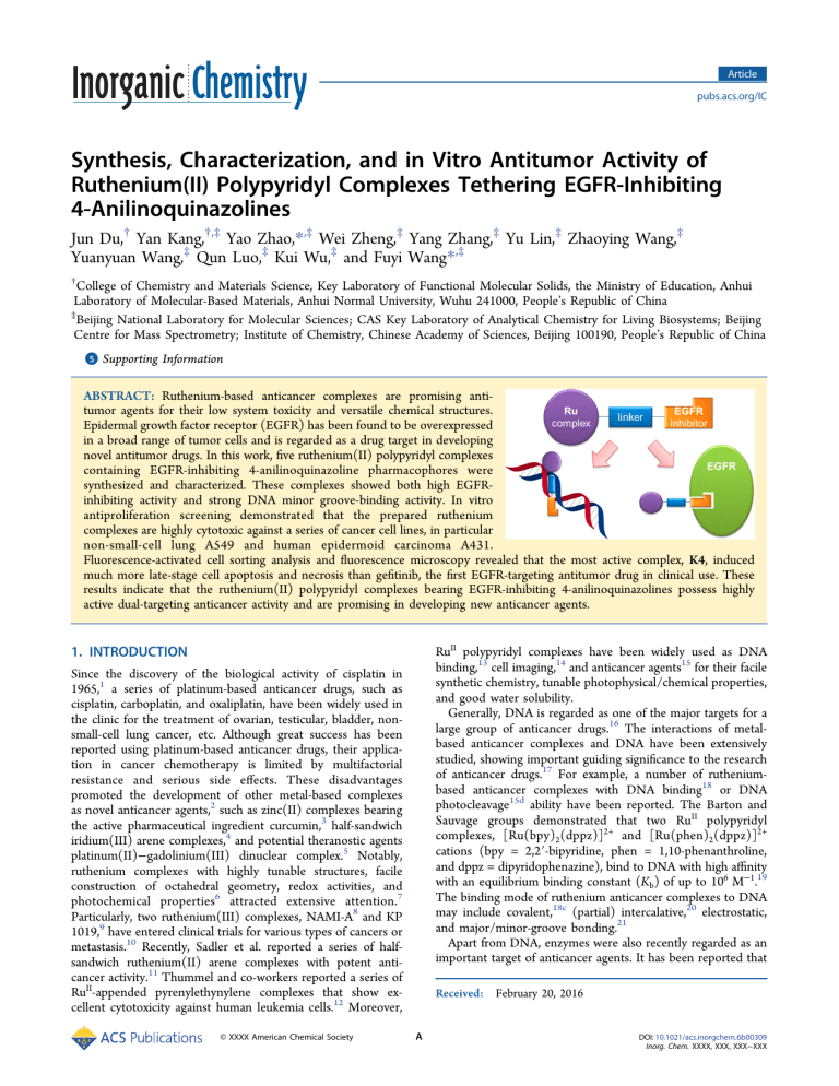
ABSTRACT: Ruthenium-based anticancer complexes are promising antitumor agents for their low system toxicity and versatile chemical structures.
Epidermal growth factor receptor (EGFR) has been found to be overexpressed
in a broad range of tumor cells and is regarded as a drug target in developing
novel antitumor drugs. In this work, five ruthenium(II) polypyridyl complexes
containing EGFR-inhibiting 4-anilinoquinazoline pharmacophores were
synthesized and characterized. These complexes showed both high EGFRinhibiting activity and strong DNA minor groove-binding activity. In vitro
antiproliferation screening demonstrated that the prepared ruthenium
complexes are highly cytotoxic against a series of cancer cell lines, in particular
non-small-cell lung A549 and human epidermoid carcinoma A431.
Fluorescence-activated cell sorting analysis and fluorescence microscopy revealed that the most active complex, K4, induced
much more late-stage cell apoptosis and necrosis than gefitinib, the first EGFR-targeting antitumor drug in clinical use. These
results indicate that the ruthenium(II) polypyridyl complexes bearing EGFR-inhibiting 4-anilinoquinazolines possess highly
active dual-targeting anticancer activity and are promising in developing new anticancer agents.
RuII polypyridyl complexes have been widely used as DNA
binding,13 cell imaging,14 and anticancer agents15 for their facile
synthetic chemistry, tunable photophysical/chemical properties,
and good water solubility.
Generally, DNA is regarded as one of the major targets for a
large group of anticancer drugs.16 The interactions of metalbased anticancer complexes and DNA have been extensively
studied, showing important guiding significance to the research
of anticancer drugs.17 For example, a number of rutheniumbased anticancer complexes with DNA binding18 or DNA
photocleavage15d ability have been reported. The Barton and
Sauvage groups demonstrated that two RuII polypyridyl
complexes, [Ru(bpy)2(dppz)]2+ and [Ru(phen)2(dppz)]2+
cations (bpy = 2,2′-bipyridine, phen = 1,10-phenanthroline,
and dppz = dipyridophenazine), bind to DNA with high affinity
with an equilibrium binding constant (Kb) of up to 106 M−1.19
The binding mode of ruthenium anticancer complexes to DNA
may include covalent,18c (partial) intercalative,20 electrostatic,
and major/minor-groove bonding.21
Apart from DNA, enzymes were also recently regarded as an
important target of anticancer agents. It has been reported that
1. INTRODUCTION
Since the discovery of the biological activity of cisplatin in
1965,1 a series of platinum-based anticancer drugs, such as
cisplatin, carboplatin, and oxaliplatin, have been widely used in
the clinic for the treatment of ovarian, testicular, bladder, nonsmall-cell lung cancer, etc. Although great success has been
reported using platinum-based anticancer drugs, their application in cancer chemotherapy is limited by multifactorial
resistance and serious side effects. These disadvantages
promoted the development of other metal-based complexes
as novel anticancer agents,2 such as zinc(II) complexes bearing
the active pharmaceutical ingredient curcumin,3 half-sandwich
iridium(III) arene complexes,4 and potential theranostic agents
platinum(II)−gadolinium(III) dinuclear complex.5 Notably,
ruthenium complexes with highly tunable structures, facile
construction of octahedral geometry, redox activities, and
photochemical properties6 attracted extensive attention.7
Particularly, two ruthenium(III) complexes, NAMI-A8 and KP
1019,9 have entered clinical trials for various types of cancers or
metastasis.10 Recently, Sadler et al. reported a series of halfsandwich ruthenium(II) arene complexes with potent anticancer activity.11 Thummel and co-workers reported a series of
RuII-appended pyrenylethynylene complexes that show excellent cytotoxicity against human leukemia cells.12 Moreover,
© XXXX American Chemical Society
Received: February 20, 2016
A
DOI: 10.1021/acs.inorgchem.6b00309
Inorg. Chem. XXXX, XXX, XXX−XXX
Article
Inorganic Chemistry
Scheme 1. Synthesis of Ruthenium Complexes K1−K5
inhibition activities, in vitro antitumor activities, in vitro
induction of apoptosis, and distribution in tumor cells were
examined. The RuII-polypyridyl substrates, such as cis-[Ru(bpy)2Cl2] and cis-[Ru(phen)2Cl2], are nontoxic to cancer cells.
However, upon coupling to an EGFR-inhibiting subunit, i.e., 4anilinoquinazoline pharmacophore, these ruthenium compounds displayed strong antiproliferation activity against cancer
cells, and dual-targeting mechanisms of action were found.
gene mutations leading to overexpression or overactivation of
protein tyrosine kinase such as epidermal growth factor
receptor (EGFR) are associated with a broad range of
malignance, such as non-small-cell lung, ovarian, breast, and
squamous cell cancers.22 EGFR, a transmembrane glycoprotein,
can bind to epidermal growth factor (EGF) and is thus
activated through dimerization and autophosphorylation of
several tyrosine residues.23 Phosphorylation of the tyrosine
residues triggers the downstream signal transduction of a
number of intracellular signaling proteins, followed by the
activation of a series of physiological processes associated with
cell growth, differentiation, apoptosis, and migration.24 Thus,
EGFR and its downstream signaling cascades have been
focused on as potential targets for the development of
anticancer agents.25 In the past decades, great effort has been
made to develop novel EGFR inhibitors as new anticancer
drugs. Among those, molecules containing 4-anilinoquinazolines were found to be highly selective EGFR inhibitors and
effective anticancer drugs. This type of antitumor agent exerts
its activity by competitive insertion into the ATP-binding
pocket of EGFR. A number of 4-anilinoquinazolines derivatives,
such as gefitinib and erlotinib, have been available in the clinic
for the treatment of non-small-cell lung cancer and squamous
carcinoma.26 Unlike the traditional cytotoxic anticancer drugs,
this kind of molecular targeting agent leads to much less
toxicity toward normal tissue. However, drawbacks such as
noncurative activity are commonly unavoidable.
Since the generation of tumors has been found to be
controlled by polygenic factors, dual- or multitargeting
treatment of tumors is a promising strategy to improve the
efficacy of therapy. For instance, ruthenium anticancer
complexes with both potent enzyme-inhibiting and DNA
interaction activity have been demonstrated.27 A platinumbased multitargeting anticancer complex was also reported,
which exhibited synergistic DNA binding and anti-inflammatory activity.28 Multityrosine-kinase inhibitors such as sorafenib29 and sunitinib30 are available in the clinic. In our group, a
series of dual-targeting ruthenium arene anticancer complexes
bearing EGFR-inhibitory pharmacophores have been designed
and synthesized,31 which showed both highly inhibitory
potency against EGFR and binding affinity with DNA.
In this work, we rationally designed a series of dual-targeting
anticancer compounds by coupling EGFR-inhibitory pharmacophores, 4-anilinoquinazoline derivatives, to noncytotoxic
RuII-polypyridyl subunits. Their structures were characterized
and their hydrolysis properties, DNA interactions, EGFR
2. RESULTS AND DISCUSSION
2.1. Chemistry. The monodentate ligands containing a 4anilinoquinazoline pharmacophore and an imidazole group for
the coordination to Ru were synthesized according to the
previous report.32 The reactions between the ligands L1 and L2
with the ruthenium polypyridyl complexes cis-[Ru(bpy)2Cl2]
and cis-[Ru (phen)2Cl2] afforded complexes cis-[Ru(bpy)2(L1)Cl](PF6) (K1), cis-[Ru(bpy)2(L2)Cl](PF6) (K2), cis-[Ru(phen) 2(L1)Cl](PF 6) (K3), cis-[Ru(phen)2 (L2)Cl](PF6 )
(K4), and cis-[Ru(bpy)2(L2)2](PF6)2 (K5), respectively
(Scheme 1). Complexes K1−K4 bear one leaving group, Cl−,
and one monodentate ligand (L1 or L2), while K5 bears no
leaving group but two monodentate ligands (L2). Complexes
K1−K4 were synthesized by mixing 1 molar equiv of cis[Ru(bpy)2Cl2] or cis-[Ru(phen)2Cl2] with the corresponding
ligand L1 or L2 in absolute methanol and refluxing at 80 °C for
10 h under argon in the dark. Complex K5 was synthesized by
refluxing cis-[Ru(bpy)2Cl2] and two molar equiv of L2 in
absolute methanol at 80 °C for 15 h under argon in the dark.
Hexafluorophosphate was added to precipitate the products
with satisfactory purification.
Complexes K1−K5 were characterized by ESI-MS, 1H NMR,
and 13C NMR spectroscopy and elemental analysis. In the 1H
NMR spectra of complexes K1−K5, the resonances between
10.18 and 7.11 ppm are assignable to the aromatic protons of
the 4-(3′ -chloro-4′-fluoroanilino)-7-methoxyquinazoline, imidazole groups, and 2,2′-bipyridine (25H in all) or 1,10′phenanthroline (25H in all). The singlets ranging from 6.66 to
6.38 ppm (1H) are assignable to the NH of 4-anilinoquinazoline pharmacophores. The typical sharp singlets at 3.7−4.0 ppm
(3H) correspond to the CH3 of the 7-methoxy group of the
quinazoline. Complexes K1−K5 have a bis-bipyridine/phenanthroline-coordinated ruthenium moiety conjugated to a derived
quinazoline group (two for K5) with a flexible C2/C3 chain.
The 4-(3′-chloro-4′-fluoroanilino)-7-methoxyquinazoline is the
active site of gefitinib, which can target the ATP-binding site of
B
DOI: 10.1021/acs.inorgchem.6b00309
Inorg. Chem. XXXX, XXX, XXX−XXX
Article
Inorganic Chemistry
Figure 1. X-ray crystal structure of complex K5. The hydrogen atoms, solvents, and PF6− anions are omitted for clarity.
chromatogram of HPLC for complex K5 showed no changes
under the same conditions, indicating the hydrolytic inertness
of K5 in PBS (Figure S5). The clean HPLC chromatograms
verify the purity of complexes K1−K5 (>90%). The stable
bonding of the ligands L1 and L2 maintained the EGFRinhibiting unit and the ruthenium polypyridyl unit of the
complexes, which validates the molecule structures in the
following experiments for biological activities.
The hydrolysis reactions of K1−K4 in PBS at 310 K were
followed by UV−vis spectroscopy, as shown in Figure 2. The
time-dependent changes in the absorbance at selected wavelength of each complex were fitted according to the first-order
reaction kinetics to give the hydrolysis rate constants (k) and
half-reaction times (t1/2), as shown in Table 1. The t1/2 of K1−
K4 ranges from 9 to 44 min, which are on the same level with
the ruthenium arene anticancer complexes bearing EGFR
inhibitor pharmacophores (11−33 min).31a It is notable that
K2 hydrolyzes faster than K1, and K4 is faster than K3,
indicating that the bulky 4-anilinoquinazoline group gives rise
to steric hindrance to the substitution of the leaving group, and
a longer linker can partially counteract the hindrance.
Moreover, the hydrolysis rate of K3 is slower than that of
K1, and K4 is slower than K2, suggesting that the steric
hindrance of the {Ru(phen)2} moiety is higher than that of
{Ru(bpy)2} imposing on the chloride group. The complexes
K1−K4 generally hydrolyze very fast, so the biological activity
research should refer to their hydrolyzed form.
2.3. EGFR Inhibition Activities. The inhibitory activities of
the ruthenium complexes K1−K5 and their ligands L1 and L2
toward EGFR were characterized by the enzyme-linked
immunosorbent assay (ELISA). The clinically available EGFR
inhibitor gefitinib was applied as a reference, with an IC50 (halfmaximal enzyme activity inhibitory concentration) value of 90
nM. The IC50 values of the Ru complexes in this work against
EGFR are listed in Table 2, and the concentration-dependent
inhibitory curves of K2, K4, and K5 are shown in Figure S6. K1
and K3 did not pass the preliminary screening, as their IC50
values are above 500 nM; thus their curves are not shown.
Ligands L1 and L2 are highly potent EGFR inhibitors, as they
keep the active site of gefitinib and do not have a steric
hindering Ru polypyridyl group. The inhibitory potency of K2
EGFR, offering molecules with potential EGFR-inhibiting
activity.
Slow diffusion of diethyl ether into a methanol solution of
K5 gave rise to red plate crystals suitable for X-ray diffraction
analysis. The X-ray structure and atom numbering are shown in
Figure 1; the crystallographic data and the selected bond
lengths, angles, and torsion angles are listed in Tables S1 and
S2 in the Supporting Information, respectively. As expected, the
ruthenium(II) center adopts a distorted octahedral geometry.
The bond angles of the bidentate bipyridine ligands with Ru
(N(1)−Ru(1)−N(2) and N(3)−Ru(1)−N(4)) are both 79°,
smaller than the angle of Ru with two monodentate-derived
imidazole ligands in L2 (N(5)−Ru(1)−N(10) = 90°). The
bond lengths between Ru and bipyridine or imidazole Ndonors are 2.04−2.09 Å, which are very close to the previously
reported bond lengths of Ru and N-donors.31c Three flexible
methylene groups between the quinazoline and the imidazole
groups make the 4-anilinoquinazoline pharmacophore and the
Ru subunit independent from each other to exert their
functions.
2.2. Hydrolysis. Ruthenium complexes containing leaving
groups, e.g., chloride, may hydrolyze under physiological
conditions. The hydrolysis may affect their chemical properties
and interactions with biological molecules. As the derived
gefitinib group is crucial for the EGFR-inhibiting activity, the
hydrolysis and ligand stability are very important for the
complexes in this work. Therefore, HPLC-MS and UV−vis
spectroscopy were employed to study the hydrolysis of K1−K5.
The hydrolysis products of the ruthenium complexes K1−K5
were analyzed by HPLC coupled mass spectrometry. The
complexes were incubated in PBS (pH = 7.4, [Cl] = 139.7
mM) at 310 K. After 1 h, an aliquot of the solution was
analyzed by HPLC-MS. The HPLC chromatograms and mass
spectra for the hydrolysis of K1−K4 are provided in the
Supporting Information (Figures S1−S4). Hydrolyzed products
with the loss of a Cl− group for K1−K4 were found. For
example, in Figure S4, the HPLC chromatogram of hydrolyzed
K4 displays two signals at 15.5 and 18.2 min, which were
identified by ESI-MS to be the aqua adduct of K4 (K4−H2O)
and intact K4, respectively. No loss of the derived 4anilinoquinazoline group (L1 or L2) was found. The
C
DOI: 10.1021/acs.inorgchem.6b00309
Inorg. Chem. XXXX, XXX, XXX−XXX
Article
Inorganic Chemistry
is higher than that of K1, and similarly, K4 is higher than K3,
which indicates that a longer linker between the 4anilinoquinazoline moiety and the ruthenium(II) polypyridyl
moiety leads to higher EGFR-inhibitory efficiency. These
results are in accordance with our previous work,31a suggesting
that a longer and flexible linker between the ruthenium moiety
and the EGFR-inhibiting pharmacophore can lower the steric
hindrance for the binding affinity to EGFR. In addition,
complex K5, bearing two EGFR-inhibiting 4-anilinoquinazoline
derivatives, shows the highest activity among K1−K5, while the
EGFR inhibitory efficiencies of complexes K2 and K4 are
comparable, indicating that, with the same alkyl linker, the
different Ru-polypyridyl moieties caused little difference in
EGFR-inhibitory activity.
2.4. Competitive DNA-Binding Assays. Ruthenium(II)
complexes with distorted octahedral structure are extensively
documented to interact with DNA via noncovalent binding,
and their cytotoxicity is usually considered to be related to their
ability to bind to DNA.18b Therefore, studies on the DNAbinding mode of ruthenium antitumor complexes are of great
importance. Hence complex K4, with the highest overall
cytotoxicity, and K5, which is structurally different from K1−
K4, are chosen to explore the binding affinity with calf thymus
DNA (ctDNA).
Various well-established DNA dyes are employed to decipher
the interaction modes of drug and DNA. Ethidium bromide
(EB), with a planar structure, is a sensitive fluorescent probe
that binds to DNA intercalatively. In aqueous solution, the
fluorescent emission of EB is quenched by water molecules,
whereas it can be greatly restored upon intercalation within
DNA base pairs. However, when another compound
competitively replaces EB from the DNA duplex, the
fluorescence intensity of the EB−DNA complex may drop,
and the binding mode of the new compound toward DNA can
be expected to be in the same way as EB.33
In this work, first EB was added to the Tris buffer solution
(pH = 7.4) of ctDNA, and bright fluorescence at ca. 587 nm
was observed. Upon adding complex K4 to the mixture, the
fluorescence intensity centered at ca. 587 nm quenched
significantly, as shown in Figure 3a. Similar quenching of
fluorescence was observed upon the addition of K5 to the
Figure 2. Kinetic study on hydrolysis of complexes K1−K4. (a, c, e, g)
Time-dependent UV−vis absorption spectra for the hydrolysis of K1−
K4 (0.01 mM) at 310 K in PBS (pH = 7.4). (b, d, f, h) Timedependent absorbance at selected wavelength for the hydrolysis of
K1−K4 (0.01 mM) at 310 K in PBS (pH = 7.4). The lines are the
fittings according to the first-order reaction kinetics.
Table 1. Hydrolysis Rate Constants (k) and Half-Times
(t1/2) of Complexes K1−K4
−4
−1
k (×10 s )
t1/2 (min)
K1
K2
K3
K4
10.3 ± 0.4
11.3 ± 0.4
13.4 ± 0.6
8.6 ± 0.4
2.6 ± 0.3
43.8 ± 0.9
7.3 ± 0.5
15.9 ± 1.2
Table 2. IC50 Values of L1 and L2 and K1−K5 for the Inhibition of EGFR Activity and of the Growth of Selected Cancer Cell
Lines
IC50 values
A549 (μM)b
EGFR (nM)a
K1
K2
K3
K4
K5
L1
L2
cisplatin
gefitinib
cis-[Ru(bpy)2Cl2]15e
cis-[Ru(phen)2Cl2]15e
>500
371
>500
254
71.6
57.4
69.6
−c
94
−
−
− EGF
40
34
34
25
23
12
18
10
16
−
−
±
±
±
±
±
±
±
±
±
4
2
6
1
2
1
2
1
1
MCF-7 (μM)b
+ EGF
27
28
15
8.6
13
13
13
−
11
−
−
±
±
±
±
±
±
±
1
0.4
2
1
2
2
2
±1
− EGF
31
35
30
12
47
35
34
13
37
>200
>200
±
±
±
±
±
±
±
±
±
HeLa (μM)b
+ EGF
4
3
3
1
2
2
2
1
1
51
33
18
14
28
28
35
−
23
−
−
±
±
±
±
±
±
±
1
1
1
1
3
3
2
±4
− EGF
74
26
42
11
50
19
47
12
15
>200
>200
±
±
±
±
±
±
±
±
±
A431 (μM)b
+ EGF
9
2
7
1
8
1
1
1
1
42
28
21
24
37
16
28
−
18
−
−
±
±
±
±
±
±
±
2
3
6
8
1
1
2
±2
− EGF
38
18
23
11
29
19
20
5.9
−
−
−
±
±
±
±
±
±
±
±
6
1
0.1
1
7
4
1
0.4
+ EGF
32
16
16
13
27
13
15
−
18
−
−
±
±
±
±
±
±
±
1
1
2
1
2
4
2
±2
The IC50 values were determined in the presence of 200 μM ATP and are the average of three independent experiments and expressed as mean ±
SD. bThe cancer cell lines were incubated with each complex for 48 h, and the IC50 values are the average of six independent experiments and
expressed as mean ± SD. c− = not tested.
a
D
DOI: 10.1021/acs.inorgchem.6b00309
Inorg. Chem. XXXX, XXX, XXX−XXX
Article
Inorganic Chemistry
enhance the effect of the EGFR inhibitors. Therefore, in this
work, the antiproliferation assay was carried out both in the
absence and in the presence of 100 ng mL−1 EGF with 48 h
incubation time after addition of the tested ruthenium(II)
complexes. In this assay, the clinically available EGFR inhibitor
gefitinib37 and the cytotoxic compound cisplatin were used as
positive controls. The IC50 values of the tested complexes are
shown in Table 2.
All the synthesized ligands and complexes exhibited
significant antiproliferation activity against the tested tumor
cell lines. As expected, adding EGF reduced their IC50 values, in
other words, increased the inhibiting potency in almost all
cases, except that for complex K4 against HeLa cells. This
suggests that EGF stimulated the repression of EGFR, which is
indeed a target of the Ru complexes in this work. Ligands L1
and L2 exhibited overall antiproliferation activity against the
tested tumor cell lines similar to gefitinib, simply because they
share the same ATP-binding site, and the structure change on
the other side is not substantial. Only complex K4 showed
increased overall potency than its corresponding ligand L2,
whereas complexes K1−K3 and K5 did not. Importantly, the
cytotoxicity of K1 and K3 against MCF-7 or HeLa cells was
much stronger than that of their corresponding Ru precursor
cis-[Ru(bpy)2Cl2].15e Similarly, K2 and K4 are much more
cytotoxic than cis-[Ru(phen)2Cl2].15e These results indicate
that a synergistic effect may be achieved by coupling the
ruthenium moieties with the EGFR-targeting groups.
Among the Ru complexes studied herein, K4 shows the
highest overall antiproliferation activity against the tumor cell
lines tested in this work, while K1 showed the lowest. Notably,
the IC50 values of K4 were equivalent to that of cisplatin against
HeLa or MCF-7 cell lines in the absence of EGF (Table 2). In
the presence of EGF, the IC50 values of K4 against A549, MCF7, and A431 cells were 8.6, 14, and 13 μM, respectively, which
are lower than those of gefitinib, 11, 23, and 18 μM,
respectively. In some cases, K2, K3, and K5 also exhibited
strong inhibition potency against the cancer cell lines. For
example, toward MCF-7 cells in the presence of EGF, the IC50
values of K3 and K5 are 18 and 28 μM, respectively, which are
comparable to that of gefitinib (23 μM). In the presence of
EGF, the IC50 values of K2 and K3 against A431 cells are both
16 μM, which are also comparable to that of gefitinib (18 μM).
Moreover, the overall antiproliferation activity of K2 or K4 is
higher than that of K1 or K3, respectively, but K1 and K2 or
K3 and K4 shared the same Ru moiety and EGFR-targeting
group. The only difference between K1 and K2 or K3 and K4 is
the length of the Cn linker (n = 2 for K1 and K3, and n = 3 for
K2 and K4). This trend is consistent with their EGFR
inhibition activity. These results again indicate that a longer
linker between the 4-anilinoquinazoline moiety and the
ruthenium(II) polypyridyl moiety can lower the steric
hindrance during their interaction with their biological targets,
which may contribute to their higher anticancer activities.
The hydrolysis rate and the biological activity for complexes
K1−K4 are controversial. On one hand, the overall activity of
K2 or K4 is higher than that of K1 or K3, respectively, in
accordance with the higher hydrolysis rate of K2 than K1 and
K4 than K3, respectively. On the other hand, although K1 or
K2 hydrolyzes faster than K3 or K4, respectively, the overall
biological activities of K3 or K4 are higher than that of K1 or
K2, respectively. Therefore, it is suggested that the hydrolysis
rate is not the only factor to determine the biological activity,
Figure 3. (a, b) Fluorescence titration of the EB−ctDNA complex
with K4 (a) or K5 (b), λex = 525 nm. (c, d) Fluorescence titration of
the Hoechst33342−ctDNA complex with K4 (c) or K5 (d), λex = 370
nm. The insets are the corresponding Stern−Volmer plots for the
quenching of fluorescence intensity upon the addition of K4 or K5.
ctDNA−EB mixture (Figure 3b), suggesting that both K4 and
K5 could replace EB and intercalatively bind to ctDNA.
Another fluorescent probe, Hoechst33342, which binds to
DNA through minor groove, was used to further examine the
binding mode of the Ru complexes in this work. Because of the
quenching by the solvent molecules, Hoechst33342 shows
weak fluorescence in Tris buffer solution (pH = 7.4); however,
upon binding to ctDNA, the fluorescence intensity at ca. 488
nm increased substantially.34 The compounds that are able to
decrease the fluorescence intensity of DNA−Hoechst33342
complex could be expected to bind to DNA in the minor
groove like Hoechst33342. As shown in Figure 3c, upon the
addition of K4, the fluorescence intensity of the ctDNA−
Hoechst33342 mixture quenched sharply. Similar quenching
was observed upon the addition of K5 (Figure 3d), suggesting
that both K4 and K5 could also replace Hoechst33342 and
bind to DNA via the minor groove mode.
The above results suggest that K4 and K5 could bind to
duplex DNA via both intercalative mode and minor groove
mode. To compare the binding affinity of the two modes, we
calculated the quenching constant35 (Ksv) for the fluorescence
intensity of EB or Hoechst bound to ctDNA by K4 or K5 with
the Stern−Volmer plot. As shown in the insets of Figure 3a−d,
the Ksv values of K4 for EB− and Hoechst−ctDNA complexes
are 3.8 × 10⁴ and 1.4 × 10⁵ M−1, respectively, and the latter is
about 2.5-fold higher than the former, suggesting that K4 tends
to interact with DNA via the minor groove mode. The Ksv
values of K5 for EB-/Hoechst−ctDNA are 1.6 × 10⁴ and 7.2 ×
104 M−1, respectively, suggesting that K5 also tends to interact
with DNA via the minor groove mode. In addition, it can be
concluded that the binding of K4 to DNA is stronger than that
of K5.
2.5. Antiproliferation Activity. The antiproliferation
activities of the ruthenium(II) complexes K1−K5 and their
ligands L1 and L2 were evaluated against four human cancer
cell lines, i.e., non-small-cell lung (A549), cervical (HeLa),
breast (MCF-7), and squamous (A431) cancer cell lines, by
means of the MTT ([3-(4,5-dimethylthiazol-2-yl)-2,5-tetrazolium bromide]) assay. These cancer cells have been reported to
overexpress EGFR,36 and EGF activates this receptor to
stimulate the fast growth of the cells and, as a consequence,
E
DOI: 10.1021/acs.inorgchem.6b00309
Inorg. Chem. XXXX, XXX, XXX−XXX
Article
Inorganic Chemistry
This result indicates that DNA may also be the target of K4,
which leads to the apoptosis of cancer cells.
Fluorescence-activated cell sorting (FACS) analysis by flow
cytometry revealed that complex K4 (Figure 4d) induced
mainly late-stage apoptosis and necrosis and showed much
more overall cytotoxicity against A549 than gefitinib (Figure
4c). This result suggests that the coupling of ruthenium(II)
polypyridyl precursor cis-[Ru(phen)2Cl2] and the EGFRinhibiting 4-anilinoquinazoline pharmacophore offers extraordinary in vitro cytotoxicity toward cancer cell line A549.
Furthermore, a positive synergistic effect may be generated
via the dual-targeting strategy.
2.7. Distribution of Ruthenium Compounds in HeLa
Cells. Time-of-flight secondary mass spectrometry (ToFSIMS) imaging was employed to analyze the cellular
distribution of Ru compounds in cancer cells. The distributions
of Ru complexes K2−K4 in single HeLa cells visualized by
ToF-SIMS imaging are shown in Figure 5. The green color
which is also influenced by other factors such as the polypyridyl
ligands.
Finally, K5, bearing two EGFR-inhibiting 4-anilinoquinazoline pharmacophores, exhibited moderate antiproliferation
activity in the absence of EGF against the cancer cells tested
compared to K1−K4. However, in the presence of 100 ng/mL
EGF, a substantial decrease of the IC50 values was observed
toward all the cancer cells tested. Considering the excellent
EGFR inhibitory activity of K5, even better than gefitinib, it is
speculated that K5 exerts its antiproliferative activities mainly
through EGFR inhibition. Similarly, L1 and L2 are also very
potent EGFR inhibitors, and their antiproliferation activities are
substantially increased upon adding EGF; their activities are
also performed through EGFR inhibition.
The Ru complexes in this work displayed higher overall
antiproliferation activity than the multitargeting organometallic
Ru complexes published previously by our group.31 However, a
dual-targeting platinum complex composed of oxoplatin and
aspirin showed more potent anticancer activity against HeLa,
MCF-7, HepG2, and A549 cells and, moreover, a lower
resistance factor for the A549 cisplatin resistance subcell line.28
2.6. Apoptosis Analysis. Since complex K4 shows the
highest overall cytotoxicity against the tested cancer cell lines
among all the ruthenium complexes in this work, its ability to
induce apoptosis was further explored. Fluorescence microscopy imaging of A549 cells incubated with first complex 4 and
then the nuclear staining dye Hoechst33342 was carried out to
evaluate the capacity of K4 to induce apoptotic cell death. As a
control in this test, gefitinib is a monofunctional EGFR
inhibitor, which exerts its effect mainly by blocking the
signaling pathway invoked by autophosphorylation of the
EGFR,26 and is expected to have less capacity to induce
apoptotic cell death. As observed from the microscopic images
(Figure 4a,b), complex K4 leads to more apoptotic bodies of
A549, characterized by the fragmentation of nuclei with
condensed chromatin, than those resulting from gefitinib.
Figure 5. TOF-SIMS images obtained from HeLa cells treated with
complex K2, K3, or K4 (50 μM). The first column depicts the sum of
signals of the 1−10 slices, which corresponds to the surficial level of
the cells (membrane), using endogenous phosphocholine fragments at
m/z 184.51 as the marker (red color). The second column
corresponds to the 51−100 slices, which depicts the deep interior of
the cells (nucleus), using endogenous deoxyribose fragments at m/z
81.26 as the marker (red color). The green color indicates the total
signal intensity of the Ru-containing fragments for K2, m/z 412.91
([K2 − L1 − HCl − PF6]+, C20H15N4O2Ru requires 413.04) and
449.65 ([K2 − L1 − PF6]+, C24H15N4O2Ru requires 449.01); K3, m/z
460.92 ([K3 − L1 − HCl − PF6]+, C24H15N4O2Ru requires 461.04)
and 497.66 ([K3 − L1 − PF6]+, C24H16N4O2ClRu requires 497.01;
K4, m/z 460.89 ([K4 − L2 − HCl − PF6]+, C24H15N4O2Ru requires
461.04) and 497.75 ([K4 − L2 − PF6]+, C24H16N4O2ClRu requires
497.01).
shows the Ru-containing fragment signals of complexes K2−K4
in all the images. The first column depicts the sum of signals of
the 1−10 slices, where the red color displays the images of the
positive ions at m/z 184, which are the endogenous
phosphocholine fragments as a marker of the cell membrane.
The second column depicts the sum of signals of the 50−100
slices, where the red color displays the images of the positive
ions at m/z 81, which are the endogenous deoxyribose
Figure 4. Confocal fluorescent images (left column, a, b) λex = 405
nm, λem = 461 nm. Flow cytometric quantification (right column, c, d)
of viable (bottom left quadrant), early-stage apoptotic (bottom right
quadrant), late-stage apoptotic (top right quadrant), and necrotic (top
left quadrant) A549 cells treated with 50 μM of corresponding
complexes in the presence of 10 nM EGF at 310 K for 24 h. The
number in each quadrant indicates the respective percentages of total
cell populations. Compounds used: (a, c) gefitinib and (b, d) K4.
F
DOI: 10.1021/acs.inorgchem.6b00309
Inorg. Chem. XXXX, XXX, XXX−XXX
Article
Inorganic Chemistry
cooling to room temperature, the mixture was filtered in a vacuum and
the filtrate was collected and evaporated to give a yellow oil. Then
deionized water (50 mL) and ethyl acetate (50 mL) were added. A
light yellow solid appeared between the water phase and ethyl acetate
phase after ultrasonic vibration for 5 min and standing for 1 h, and the
mixture was filtered under vacuum and washed with water and ethyl
acetate to give L1 as light yellow powder (0.8 g, 62%). Mp: 259−261
°C. Ligand L1 is slightly soluble in DMSO-d6, but the solubility is too
low to allow for 13C NMR measurements. ESI-MS: m/z 414.2 ([M +
H]+ requires 414.1). 1H NMR (400 MHz, DMSO-d6, TMS): δH
(ppm) 9.53 (s, 1H), 8.47 (s, 1H), 8.06 (dd, J1 = 4.0 Hz, J2 = 2.4 Hz,
1H), 7.77 (s, 1H), 7.74−7.71 (m, 2H), 7.41 (t, J1 = J2 = 9.2 Hz, 1H),
7.29 (s, 1H), 7.19 (s, 1H), 6.91 (s, 1H), 4.48 (t, J1 = J2 = 5.2 Hz, 2H),
4.38 (t, J1 = 4.8 Hz, J2 = 5.6 Hz, 2H), 3.93 (s, 3H). Anal. (%) Calcd for
C20H17ClFN5O2: C, 58.05; H, 4.14; N, 16.9. Found: C, 58.04; H, 4.20;
N, 16.32.
6-(2-(3-(1H-Imidazol-1-yl) propoxy)-4-(3′-chloro-4′-fluoroanilino)-7-methoxyquinazoline (L2). 4-(3′-Chloro-4′-fluoroanilino)-6-hydroxy-7-methoxyquinazoline (12.0 mg, 37.6 mmol) and potassium
carbonate (24.0 mg, 173.6 mmol) were mixed in acetone (500 mL).
Then 1,3-dibromopropane (15.2 mL, 149.2 mmol) was added, and the
resulting mixture was refluxed for 10 h. After cooling to room
temperature, the mixture was filtered under vacuum and the filtrate
was collected. Then the solvent was evaporated under vacuum, and the
residue was recrystallized from ethanol. The yellow residue was further
purified by flash chromatography on silica gel using ethyl acetate/
petroleum (3:1) as eluent to give 4-(3′-chloro-4′-fluoroanilino)-6-(2bromopropoxy)-7-methoxyquinazoline (L2′) as white powder (6.0 g,
36%). Imidazole (516 mg, 7.6 mmol), TBAB, (60 mg, 0.2 mmol), and
NaOH (s) (908 mg, 22.6 mmol) were mixed in acetonitrile (100 mL).
The reaction mixture was heated to 90 °C and refluxed for 1 h; then
compound L2′ (2.0 g, 4.54 mmol) was added and the mixture was
refluxed for 5 h. After cooling to room temperature, the mixture was
filtered under vacuum and the filtrate was collected and evaporated to
give a yellow oil. Then water (80 mL) and ethyl acetate (80 mL) were
added. A light yellow solid appeared between the water phase and the
ethyl acetate phase after ultrasonic vibration for 5 min and standing for
1 h, and the mixture was filtered under vacuum and washed with water
and ethyl acetate to give L2 as a light yellow powder (1.4 g, 72%). Mp:
201−203 °C. ESI-MS: m/z 428.2 ([M + H]+ requires 428.1). 1H
NMR (400 MHz, DMSO-d6, TMS): δH (ppm) 9.51 (s, 1H), 8.50 (s,
1H), 8.10 (dd, J1 = 2.4 Hz, J2 = 2.8 Hz, 1H), 7.79−7.75 (m, 2H), 7.64
(s, 1H), 7.43 (t, J1 = J2 = 9.2 Hz, 1H), 7.22 (s, 2H), 6.91 (s, 1H), 4.20
(t, J1 = J2 = 6.8 Hz, 2H), 4.08 (t, J1 = J2 = 6.0 Hz, 2H), 3.97(s, 3H),
2.33−2.27(m, 2H). 13C NMR (DMSO-d6,400 MHz, TMS): δC (ppm)
156.49, 155.01, 153.20, 148.51, 147.61, 137.84, 129.05, 123.91, 122.70,
119.84, 119.33, 119.15, 117.04, 116.83, 109.20, 107.88, 103.47, 66.17,
56.41, 43.39, 30.55. Anal. (%) Calcd for C21H19ClFN5O2·H2O: C,
56.57; H, 4.75; N, 15.71. Found: C, 56.51; H, 4.61; N, 15.46.
Synthesis and Characterization of [(N∧N)2Ru(L)Cl]PF6 (K1−
K5). General Procedure. The five complexes K1−K5 were prepared
following the methods below. The 4-anilinoquinazoline derivative L1
or L2 (0.1 mmol) and corresponding cis-[(N∧N)2Ru(Cl)2] (cis[Ru(bpy)2Cl2] or cis-[Ru(phen)2Cl2], 0.1 mmol for K1−K4, 0.2 mmol
for K5) were dissolved in methanol (60 mL), and the mixture was
refluxed under Ar in the dark until the solution became clear. After
cooling to room temperature, the solution was filtered and excess
ammonium hexafluorophosphate (0.3 mmol for K1−K4, 0.4 mmol for
K5) was added to this mixture and further stirred for 2 h at 318 K to
precipitate the product. The solid collected after filtration was washed
with excess methanol and recrystallized from MeOH to give the
product. Complexes K1−K5 were characterized by ESI-MS, 1H NMR
spectroscopy, 13C NMR spectroscopy, and elemental analysis, as
shown below.
K1: MS (m/z): 431.571 ([M − PF6 + H]2+ requires 431.562). 1H
NMR (DMSO-d6, 400 MHz): δ (ppm) 9.86 (d, J = 5.2 Hz, 1H), 9.59
(s, 1H), 8.73 (d, J = 8 Hz, 1H), 8.64−8.61 (m, 2H), 8.56 (d, J = 8 Hz,
2H), 8.39 (d, J = 5.2 Hz, 1H), 8.10 (t, J1 = 8.4 Hz, J2 = 8 Hz, 3H), 8.04
(t, J1 = 7.6 Hz, J2 = 7.2 Hz, 1H), 7.89−7.83 (m, 3H), 7.77−7.72 (m,
3H), 7.55−7.50 (m, 2H), 7.46 (t, J1 = J2 = 9.2 Hz, 1H), 7.37 (s, 1H),
fragments as a cell nucleus marker. All the images depict the
overlay of the Ru-containing fragments and the endogenous
markers. The SIMS images of individual markers are listed in
the Supporting Information (Figures S7−S9). The results show
that complexes K2−K4 could penetrate the HeLa cell
membrane and go deep inside the cells. Complexes K2 and
K3 were located not only in the cell membrane but also in the
cell nucleus. Whereas for K4 only a small amount was found in
the membrane and the nucleus region, a large amount of K4
was found in the cytoplasm. Notably, cells treated by complex
K4 were observed to fragment, which may be due to the higher
antiproliferation activity of K4 than that of the other complexes.
Inductively coupled plasma mass spectrometry (ICP-MS)
was used to evaluate the binding affinity of Ru complexes to
DNA and the membrane proteins in cancer cells. For this
purpose, ∼107 A549 cancer cells were treated with K4 for 48 h,
and then DNA and membrane proteins were extracted,
respectively, and the level of ruthenium binding to DNA or
membrane proteins was determined by ICP-MS. The level of
K4 was 36.7 ng Ru/mg DNA and 618 ng Ru/mg of membrane
proteins. This result supports the SIMS results that complex K4
was able to accumulate in both the cell membrane and nuclei of
the A549 cancer cells, binding to membrane proteins, most
likely EGFR, and DNA. This result further verifies that complex
4 can target both membrane proteins and DNA, exhibiting
dual-targeting potential, although the Ru uptake of membrane
proteins is about 18-fold higher than that of DNA.
3. EXPERIMENTAL SECTION
Materials. RuCl3·3H2O (Ru > 36.7%) was purchased from
Shenyang Jingke Reagent Co. (China), bipyridine, phenanthrolin,
and NH4PF6 were from Alfa Aesar, DMSO, cisplatin, and trifluoroacetic acid (TFA) were from Sigma, 1,2-dibromoethane, 1,3dibromopropane, and imidazole were from Beijing Ouhe Technology
Co. (China), and 4-(3-chloro-4′-fluoroanilino)-6-hydroxy-7-methoxyquinazoline (AR grade) was from Shanghai FWD Chemicals Co.
(China). Cis-[Ru(bpy)2Cl2] and cis-[Ru(phen)2Cl2] were synthesized
following methods reported in the literature.38 Organic solvents
including absolute methanol, absolute ethanol, absolute ether,
acetonitrile, dichloromethane, and THF were all analytical grade and
used directly without further purification. The deionized water used in
the experiments was prepared by a Milli-Q system (Millipore, Milford,
MA, USA). The protein tyrosine kinase epidermal growth factor
receptor and the epidermal growth factor were purchased from Sigma,
and other biological agents including the ELISA kits for EGFR
inhibitor screening were from Cell Signaling Technology Inc. (USA).
1
H NMR and 13C NMR were recorded on an Avance III 400
spectrometer (Bruker) at 400 MHz for 1H and 100.6 MHz for 13C.
Synthesis and Characterization. 6-(2-(2-(1H-Imidazol-1-yl)ethoxy)-4-(3′-chloro-4′-fluoroanilino)-7-methoxyquinazoline (L1).
Compound L1 was synthesized following a method reported in the
literature31a,32 with minor modifications. 4-(3′-Chloro-4′-fluoroanilino)-6-hydroxy-7-methoxyquinazoline (8.0 g, 25 mmol) and potassium
carbonate (18.0 g, 115.9 mmol) were mixed in DMF (300 mL). Then
1,2-dibromoethane (8 mL, 92.4 mmol) was added, and the resulting
mixture was heated at 80 °C for 8 h. After cooling to room
temperature, the mixture was filtered under vacuum, and the filtrate
was collected. Then the solvent was evaporated in a vacuum, and the
residue was recrystallized from ethanol. The yellow crude product was
further purified by flash chromatography on silica gel using ethyl
acetate/petroleum (5:2) as eluent to give 4-(3′-chloro-4′-fluoroanilino)-6-(2-bromoethoxy)-7-methoxyquinazoline (L1′) as white powder
(4.8 g, 45%). Imidazole (355 mg, 5.2 mmol), tetrabutyl ammonium
bromide (TBAB) (41.3 mg, 0.133 mmol), and NaOH (s) (624 mg,
15.6 mmol) were mixed in acetonitrile (60 mL). The reaction mixture
was heated to 90 °C and refluxed for 1 h; then compound L1′ (1.33 g,
3.12 mmol) was added, and the mixture was refluxed for 5 h. After
G
DOI: 10.1021/acs.inorgchem.6b00309
Inorg. Chem. XXXX, XXX, XXX−XXX
Article
Inorganic Chemistry
7.28 (dd, J1 = J2 = 7.2 Hz, 2H), 7.23 (s, 1H), 6.41 (s, 1H), 4.54 (t, J1 =
J2 = 5.6 Hz, 2H), 4.36 (t, J1 = J2 = 3.6 Hz, 2H), 3.88 (s, 3H). Anal. (%)
Calcd for C40H37Cl2F7N9O4PRu (M + 2H2O): C, 46.03; H, 3.57; N,
12.08. Found: C, 46.06; H, 3.35; N, 12.29.
K2: MS (m/z): 420.591 ([M − PF6 − Cl]2+ requires 420.582). 1H
NMR (MeOD, 400 MHz): δ (ppm) 9.81 (d, J = 5.2 Hz, 1H), 8.44−
8.36 (m, 4H), 8.30 (dd, J1 = J2 = 8 Hz, 2H), 8.11 (s, 1H), 7.97 (d, J =
1.2 Hz, 1H), 7.89−7.82 (m, 2H), 7.77−7.67 (m, 5H), 7.62 (s, 1H),
7.59(d, J = 5.6 Hz, 1H), 7.49 (t, J1 = J2 = 6.4 Hz, 1H), 7.35 (t, J1 = J2 =
6.4 Hz, 1H), 7.20−7.11 (m, 5H), 6.44 (s, 1H), 4.32−4.23 (m, 2H),
3.97 (m, 3H), 3.93−3.86 (m, 2H), 2.28−2.20 (m, 2H). Anal. (%)
Calcd for C41H40Cl2F7N9O4.5PRu (M + 2.5H2O): C, 46.16; H, 3.78;
N, 11.82. Found: C, 46.11; H, 3.77; N, 11.84.
K3: MALDI-TOF-MS (m/z): 437.576 ([M − PF6 − Cl]2+ requires
437.574). 1H NMR (DMSO-d6, 400 MHz): δ (ppm) 10.18 (d, J = 4.8
Hz, 1H), 9.43 (s, 1H), 8.85 (d, J = 4.8 Hz, 1H), 8.71(dd, J1 = 8 Hz, J2
= 8.4 Hz, 2H), 8.52 (s, 1H), 8.40 (dd, J1 = J2 = 8 Hz, 2H), 8.31 (d, J =
8.8 Hz, 1H), 8.25−8.13 (m, 5 H), 8.07 (dd, J1 = J2 = 2.4 Hz, 1H), 8.00
(d, J = 5.2 Hz, 1H), 7.92 (dd, J1 = J2 = 5.2 Hz, 1H), 7.76−7.72 (m,
3H), 7.49−7.39 (m, 3H), 7.29 (s, 1H), 7.21 (s, 1H), 6.44 (s, 1H),
4.52−4.45 (m, 2H), 4.36−4.27 (m, 2H), 3.83 (s, 3H). Anal. (%) Calcd
for C44H36Cl2F7N9O3.5PRu (M + 1.5H2O): C, 48.81; H, 3.35; N,
11.64. Found: C, 48.98; H, 3.36; N, 11.38.
K4: MALDI-TOF-MS (m/z): 462.595 ([M − PF6 + H]2+ requires
462.571). 1H NMR (DMSO-d6, 400 MHz): δ (ppm) 10.04 (d, J = 4.8
Hz, 1H), 9.43 (s, 1H), 8.81 (d, J = 4.9 Hz, 1H), 8.62 (d, J = 8.0 Hz,
1H), 8.54 (d, J = 8.0 Hz, 1H), 8.50 (s, 1H), 8.34 (d, J = 7.9 Hz, 1H),
8.28−8.23 (m, 2H), 8.12 (t, J1 = J2 = 8.4 Hz, 2H), 8.06−7.99 (m, 3H),
7.94−7.92 (m, 2H), 7.90−7.88 (m, 1H), 7.74−7.70 (m, 1H), 7.65 (d, J
= 6.4 Hz, 2H), 7.40−7.36 (m, 3H), 7.22 (s, 1H), 7.16 (s, 1H), 6.46 (s,
1H), 4.09 (t, J1 = 6.8 Hz, J2 = 6.4 Hz, 2H), 3.88−3.83 (m, 5H), 2.17−
2.11(m, 2H). Anal. (%) Calcd for C45H39Cl2F7N9O4PRu (M + 2H2O):
C, 48.88; H, 3.55; N, 11.40. Found: C, 49.02; H, 3.40; N, 11.36.
K5: MALDI-TOF-MS (m/z): 634.138 ([M − 2PF6]2+ requires
634.143). 1H NMR (DMSO-d6, 400 MHz): δ (ppm) 9.43 (s, 2H),
8.85 (d, J = 5.2 Hz, 2H), 8.45 (d, J = 8 Hz, 4H), 8.35 (d, J = 8.2 Hz,
2H), 8.04 (dd, J1 = J2 = 2.4 Hz, 2H), 7.94 (t, J1 = J2 = 7.6 Hz, 2H),
7.77−7.69 (m, 8 H), 7.64 (s, 2H), 7.59 (t, J1 = J2 = 6.4 Hz, 2H), 7.39
(t, J1 = J2 = 8.8 Hz, 2H), 7.29−7.25 (m, 4H), 7.16 (s, 2H), 6.66 (s,
2H), 4.05 (t, J1 = J2 = 6.8 Hz, 4H), 3.90−3.86 (m, 4H), 3.83 (s, 6H),
2.14−2.11 (m, 4H). Anal. (%) Calcd for C62H58Cl2F14N14O6P2Ru (M
+ 2H2O): C, 46.68; H, 3.66; N, 12.29. Found: C, 46.72; H, 3.47; N,
12.20.
X-ray Crystallography. A crystal of K5 suitable for single-crystal
X-ray diffraction with a size of 0.21 × 0.17 × 0.04 mm3 was selected.
Data were collected on an MM007-HF CCD (Saturn 724+)
diffractometer in ω scans with confocal-monochromated Mo Kα (λ
= 0.710 73 Å) radiation. The structure was refined with full-matrix
least-squares on F2 using the SHELXL (Sheldrick, 2013) programs.
Crystal parameters and details of the data collection and refinement
are shown in Table S1. Selected bond lengths (Å) and bond angles (°)
are shown in Table S2.
Electrospray Ionization Mass Spectroscopy (ESI-MS). The
positive-ion ESI mass spectra for the hydrolytic products were
obtained with a Xevo G2 Q-TOF (Waters USA), which was equipped
with a Masslynx (ver. 4.0) data processing system for analysis and
postprocessing. The spray voltage and the cone voltage were 3.5 kV
and 5 V, respectively. The desolvation temperature was 623 K, and the
source temperature 373 K. Nitrogen was used as both cone gas and
desolvation gas with a flow rate of 50 and 800 L h−1, respectively. The
spectra were acquired in the range 200−2000 m/z.
Hydrolysis of Complexes. The kinetic studies on the hydrolysis
of complexes K1−K5 were carried out employing a UV-2550
spectrometer (Shimadzu, Japan). First, the tested complex was
dissolved in DMSO at a concentration of 2 mM, an aliquot (15 μL)
of the DMSO solution was then added to 2985 μL of PBS (pH = 7.4)
in a quartz cuvette, and the UV−vis spectra of the mixture were
immediately recorded by scanning over the wavelength ranging from
200−800 nm at 5 min intervals at 37 °C. The wavelength
corresponding to the maximum absorbing changes of each hydrolysis
reaction was selected for measurement of the rate constant. The same
procedures as described above were used to prepare the samples for
the kinetic study. The absorbance at selected wavelength for each
complex was recorded at 5 min intervals. The time-dependent
absorbance was fitted using Origin 8.0 (OriginLab Corporation, USA)
to give the first-order rate constant k, and the half-reaction time t1/2
was calculated by the following formula:
A = C e−kt + A 0
t1/2 = ln 2/k
where A is the absorbance and A0 and C are constants.
To identify the hydrolysis products, the samples were prepared by
diluting a DMSO solution of the each complex (10 mM) with PBS
(pH = 7.4) to 1 mM. The solution was incubated at 310 K for 1 h, an
aliquot of which was then analyzed by HPLC coupled with ESI-MS.
An Agilent 1200 series quaternary pump and a Rheodyne sample
injector with a 20 μL loop, an Agilent 1200 series UV−vis DAD
detector, and Chemstation data processing system were used. The
mobile phase solvent A was water containing 0.1% TFA, and solvent B
was acetonitrile containing 0.1% TFA. The separation of hydrolytic
adducts of the ruthenium complexes was carried out on an Agilent
Eclipse XDB-C18 reversed-phase column (4.6 × 150 mm, 5 μm). The
gradient B was 10% to 60% from 0 to 25 min, 60% to 80% for 3 min,
and 80% for 2 min.
In Vitro Antiproliferation Assays. The human lung adenocarcinoma A549, human cervical cancer HeLa, human breast cancer MCF7, and human epidermoid carcinoma A431 cell lines were obtained
from the Centre for Cell Resource of Peking Union Medical College
Hospital and were maintained in DMEM medium supplemented 90%
DMEM (Invitrogen, USA) + 10% fetal bovine serum (Invitrogen,
USA) + 1% penicillin−streptomycin (Invitrogen, USA). On request,
an aliquot of 100 ng mL−1 epidermal growth factor (Sigma, USA) was
added into the media. The cells were grown at 310 K in a humidified
atmosphere containing 5% CO2 for 2−3 days prior to screening
experiments.
The IC50 values were determined by the MTT assay. Cells were
counted by a Luna automated counter (Logos Biosystems, Korea) and
were plated at a density of 5000 cells/well (A549), 6500 cells/well
(HeLa), 8000 cells/well (MCF-7), and 8000 cells/well (A431),
respectively, in 100 μL of media in 96-well plates and grew in the
absence or the presence of EGF for 24 h. The stock solutions (10 mM,
except for cisplatin (1 mM)) of all tested complexes were made up
fresh in DMSO before diluted down in media to give the required
concentration for addition to the cells. For each ruthenium complex,
eight different concentrations were prepared from the stock solution
by diluting with the cell culture medium prior to use, and the
concentration of DMSO was kept 1% in all dilutions. Cells were then
exposed to each tested complex at various concentrations for 48 h.
Then the drug media was discarded and washed three times with PBS,
and 100 μL of complete medium containing MTT (0.5 mg/mL) was
added to each well and incubated at 310 K for 4 h. The MTT media
was removed, and 100 μL of DMSO was added to each well to
dissolve the crystals at room temperature for 10 min. Optical density
(OD) for each well was measured using a microplate reader
(SpectraMax M5, Molecular Devices Corporation) at the wavelength
of 570 nm. The inhibition rate (IR) was calculated based on the
following equation:
IR(%) = [1 − (ODcomplex − ODblank )/(ODcontrol − ODblank )]
× 100%
All values of IR reported were averages of six independent
experiments and expressed as mean ± SD (standard deviation).
Competitive Displacement of DNA Binding Assays. The EB
displacement assay was done in Tris buffer solution (5 mM, pH = 7.4).
The concentration of ctDNA and EB was kept at 20 and 200 μM,
respectively, in the solution, which was titrated with varying
concentrations of K4 or K5 from 0 to 100 μM. The ctDNA-EB
complex was excited at 525 nm, and the emission spectra were
H
DOI: 10.1021/acs.inorgchem.6b00309
Inorg. Chem. XXXX, XXX, XXX−XXX
Article
Inorganic Chemistry
read on the ELISA plate reader (SpectraMax M5Molecular Devices
Corporation) at 450 nm to determine the OD values.
Confocal Microscopic Analysis. A total of 1.2 × 10⁵ A549 cells
per well were plated in a laser scanning confocal Petri dish and grown
in the absence/presence of EGF for 24 h. A 2.5 mg amount of
Hoechst33342 was dissolved in 1 mL of deionized water, then diluted
to 25 μg mL−1 by medium. After removing the cell culture medium
and washing once with PBS, 1 mL of 1 μg mL−1 Hoechst33342 was
added in the dark. After being incubated at 37 °C for 10 min, the cells
were washed three times by 1 mL of PBS. The cells were maintained
by colorless minimal medium. Fluorescence images were obtained by
an FV1000-IX81 confocal laser scanning microscope (Olympus), at an
excitation wavelength of 405 nm and emission wavelength of 425−500
nm.
Flow-Cytometry Double-Staining Assay. A549 cells were
seeded at a density of 2 × 10⁵ per well in a six-well plate and allowed
to attach for 16 h; then the cells were maintained with the
corresponding complexes at 310 K for 24 h. The supernatant was
removed, and cells were detached by trypsinization after washing with
PBS. The cells were transferred to FACS tubes after washing by PBS
and centrifuged at 1000 rpm for 3 min. After resuspension in 0.5 mL
of binding buffer, the cells were incubated with 5 μL of annexin-V
conjugate for 5 min, followed by addition of 5 μL of 7-AAD in the dark
prior to the FACS analysis. The FACS assays were performed on a
Calibur flow cytometer (BD, Franklin Lakes, NJ, USA), of which the
FL2 channel was used to record the intensity of annexin V-PE staining
and the FL3 channel to record the intensity of 7-AAD staining. The
data were quantified by Sell Quest software (BD).
Secondary Ion Mass Spectrometry Imaging. HeLa cells were
seeded on silicon wafers at a density of 1 × 10⁴ mL−1 in a cell culture
dish in the medium solution containing 90% DMEM, 10% FBS, and
1% PS and incubated at 5% CO2 and 37 °C with 50 μM tested
ruthenium complex (K2, K3, or K4) for 24 h. Control cells were
incubated alongside the ruthenium complex doped cells. The
supernatants were removed, and cells were washed three times by
ammonium acetate (150 mM, pH = 7.4). Then the cells were frozen
using liquid nitrogen and transferred intermediately into an LGJ-12
lyophilizer (Beijing Songyuanhuaxing Technology Develop Co., Ltd.)
for freeze-drying overnight. ToF-SIMS analysis was conducted on a
ToF-SIMS V mass spectrometer (IONTOF GmbH, Munster,
Germany). Dual-beam experiments were performed using a 10 keV
argon cluster ion beam (Arn+) as sputtering beam and a 30.0 keV Bi3+
beam as analysis beam. High spatial resolution images were collected
by 256 × 256 pixels with the highest resolution of 500 nm over a 100
× 100 μm2 area using a pulsed analysis beam (dc current = 200 pA,
pulse width = 23 ns, and repetition rate = 5 kHz) at the center of a 300
× 300 μm2 crater eroded by an Arn+ sputtering source. The current of
the Arn+ was ∼2 nA with a lead-off time of 60 μs. Positive ion spectra
were recorded and calibrated by H+, CH3+, and C2H5+. Signals were
collected layer by layer, and images were conducted by using IONTOF SurfaceLab software (version 6.4, ION-TOF, Mü nster,
Germany) by combining specific slice(s). The signal intensities were
displayed on a color scale, which were directly related to the level of
detected ions of interest.
Cellular Uptake Studies. Complex K4 was dissolved in DMSO to
yield a 6 mM stock solution. A549 cells were seeded in a Corning cell
culture dish containing 8 mL of growth medium. When the coverage
was over 90%, the cells were treated with 50 μM (0.5% DMSO) of the
complex at 310 K for 48 h. The cells were also incubated in intact
medium (0.5% DMSO) as controls. Then the media were removed,
and the cells were washed with PBS solution three times. PBS
containing 0.04% EDTA (4 mL) was used to detach the cells. The
combined cells were centrifuged for 2 min at 4 °C, and the cells were
washed three times with 1 mL of ice-cold PBS. The suspension was
divided into two parts. One part was used to analyze the metal content
in the membrane proteins, and the other was used for DNA-bound
ruthenium analysis. The Bestbio-Membrane protein extraction kit and
TIANamp genomic DNA kit, RNase A (Tiangen Biotech (Beijing)
Co., Ltd.), were used to extract the membrane proteins and nuclear
fractions. The concentration of extracted proteins was determined by
recorded from 535 to 800 nm. Emission spectra were obtained on a
Hitachi F-4500 fluorescence spectrophotometer (Japan). Measurement parameters: PMT voltage, 700 V; EX slit, 10.0 nm; EM slit, 10.0
nm.
The Hoechst33342 displacement assay was also done in Tris buffer
solution (5 mM, pH = 7.4). The concentration of ctDNA and
Hoechst33342 was kept at 20 and 200 μM, respectively, in the
solution, which was titrated with varying concentrations of competing
complexes from 0 to 100 μM. The ctDNA−Hoechst33342 complex
was excited at 370 nm, and emission spectra were recorded from 400
to 680 nm. Measurement parameters: PMT voltage, 700 V; EX slit, 5.0
nm; EM slit, 5.0 nm.
The Stern−Volmer constant (Ksv) was used to evaluate the
fluorescence quenching efficiency. The classical Stern−Volmer
equation is F0/F = 1 + Ksv[Q], where F0 and F are the fluorescence
intensities before and after the addition of the quencher, respectively,
[Q] is the concentration of the quencher, and Ksv is the quenching
constant.
Enzyme-Linked Immunosorbent Assay (ELISA). ELISA, a
widely used in vitro screening method for enzyme inhibitors, was
applied to characterize the inhibition potency of ruthenium(II)
polypyridyl complexes containing 4-anilinoquinazoline ligands toward
EGFR. The receptor tyrosine kinase solution in 50% glycerol,
containing 50 mM HEPES (pH = 7.6), 150 mM NaCl, 0.1% Triton,
and 1 mM dithiothreitol (DTT), was purchased from Sigma; phosphotyrosine mouse mAb (P-Tyr-100), signal transduction protein (Tyr66)
biotinylated peptide, adenosine-triphosphate (ATP), and dl-DTT
HTScan tyrosine kinase buffer (4×) were purchased from Cell
Signaling Company; HRP-labeled goat anti-mouse lgG (H + L) was
purchased from Zhongshan Golden Bridge Biotechnology Co. Ltd.
(China); bovine serum albumin (BSA) and 3,3′,5,5′-tetramethylbenzidine (TMB) were from Xinjingke Biotechnology Co. Ltd. (China);
streptavidin was from Tianjin Biotechnology Co. Ltd. (China); and
96-well plates were purchased from Beijing Bio Dee Bio Tech Co. Ltd.
The ELISA screening was performed following the instructions
provided by the supplier of the assay kits (No. 7909, Cell Signaling
Technology, Inc.). An aliquot (0.12 μL) of the enzyme solution was
added to 4.38 μL of DTT kinase buffer, which consists of 5 mM DTT
and 240 mM HEPES (pH = 7.5), 20 mM MgCl2, 20 mM MnCl2, and
12 μM Na3VO4. Each complex was dissolved in dimethyl sulfoxide
(DMSO) to give a 4 mM solution, which was diluted with 0.05%
Tween-20 in deionized water to give a 40 μM solution. The ATP/
peptide mixture was prepared by addition of 0.36 μL of 10 mM ATP
to 4.5 μL of 6 μM substrate peptide and then diluted with D2O to 9
μL.
Each well of a microtiter plate was coated with 100 μL of 10 μg
mL−1 streptavidin in carbonate−bicarbonate buffer, incubated overnight at 277 K, and then blocked with 1.5% BSA in PBS/T (PBS
solution contained 0.05% Tween-20) at 310 K for 2 h, followed by
three washings with PBS/T prior to use.
Various concentrations of tested complexes (4.5 μL) with 1%
DMSO were added to 4.36 μL of DTT/buffer and 0.12 μL of 188 ng
μL−1 EGFR and incubated at 298 K for 5 min, followed by addition of
the mixture of 0.45 μL of PTP1B (Tyr66), 0.36 μL of ATP, and 4.14
μL of D2O, and then the resulting mixture was incubated at 310 K for
1 h. The phosphorylation reaction was terminated by the addition of
18 μL/well stop buffer (50 mM EDTA, pH = 8). Then, 25 μL/well of
each enzymatic reaction mixture and 75 μL/well of D2O were added
to the plate (in triplicate) for incubation at 310 K for 1 h. Following
three washings with PBS/T, 100 μL of primary antibody (P Tyr-100,
1:1000 in PBS/T with 1.5% BSA) was added to each well, and the
plate was incubated at 310 K for another 1 h. The plate was again
washed three times with PBS/T, and then 100 μL of secondary
antibody (HRP-labeled goat anti-mouse lgG, 1:1000 in PBS/T with
1.5% BSA) was added to each well for 1 h of incubation at 310 K,
followed by three washings with PBS/T. Finally, 100 μL of TMB
substrate (TMB (1 mg mL−1): citric acid-dibasic sodium phosphate
buffer (pH = 5.0):30% H2O2 = 100:900:1) was added to each well, the
plate was incubated at 310 K for 15 min, then the reaction was stopped
by addition of 100 μL of 2 M H2SO4 to each well, and the plate was
I
DOI: 10.1021/acs.inorgchem.6b00309
Inorg. Chem. XXXX, XXX, XXX−XXX
Article
Inorganic Chemistry
(2) (a) Mjos, K. D.; Orvig, C. Chem. Rev. 2014, 114 (8), 4540−4563.
(b) Santini, C.; Pellei, M.; Gandin, V.; Porchia, M.; Tisato, F.;
Marzano, C. Chem. Rev. 2014, 114 (1), 815−862. (c) RomeroCanelón, I.; Sadler, P. J. Inorg. Chem. 2013, 52 (21), 12276−12291.
(d) Jakupec, M. A.; Galanski, M.; Arion, V. B.; Hartinger, C. G.;
Keppler, B. K. Dalton Trans. 2008, 2, 183−194.
(3) Mendiguchia, B. S.; Pucci, D.; Mastropietro, T. F.; Ghedini, M.;
Crispini, A. Dalton Trans. 2013, 42 (19), 6768−6774.
(4) Liu, Z.; Romero-Canelon, I.; Habtemariam, A.; Clarkson, G. J.;
Sadler, P. J. Organometallics 2014, 33 (19), 5324−5333.
(5) Zhou, W.; Wang, X.; Hu, M.; Zhu, C.; Guo, Z. Chem. Sci. 2014, 5
(7), 2761.
(6) Bergamo, A.; Sava, G. Chem. Soc. Rev. 2015, 44 (24), 8818−8835.
(7) Clarke, M. J. The Potential of Ruthenium in Anticancer
Pharmaceuticals. In Inorganic Chemistry in Biology and Medicine;
American Chemical Society: Washington DC, 1980; Vol. 140, pp
157−180.
(8) Groessl, M.; Reisner, E.; Hartinger, C. G.; Eichinger, R.;
Semenova, O.; Timerbaev, A. R.; Jakupec, M. A.; Arion, V. B.; Keppler,
B. K. J. Med. Chem. 2007, 50, 2185−2193.
(9) Ang, W. H.; Dyson, P. J. Eur. J. Inorg. Chem. 2006, 2006 (20),
4003−4018.
(10) (a) Rademaker-Lakhai, J. M.; van den Bongard, D.; Pluim, D.;
Beijnen, J. H.; Schellens, J. H. M. Clin. Cancer Res. 2004, 10 (11),
3717−3727. (b) Hartinger, C. G.; Zorbas-Seifried, S.; Jakupec, M. A.;
Kynast, B.; Zorbas, H.; Keppler, B. K. J. Inorg. Biochem. 2006, 100 (5−
6), 891−904.
(11) (a) Wang, F.; Chen, H.; Parsons, S.; Oswald, I. D.; Davidson, J.
E.; Sadler, P. J. Chem. - Eur. J. 2003, 9 (23), 5810−5820. (b) Yan, Y.
K.; Melchart, M.; Habtemariam, A.; Sadler, P. J. Chem. Commun. 2005,
38, 4764−4776.
(12) Lincoln, R.; Kohler, L.; Monro, S.; Yin, H.; Stephenson, M.;
Zong, R.; Chouai, A.; Dorsey, C.; Hennigar, R.; Thummel, R. P.;
McFarland, S. A. J. Am. Chem. Soc. 2013, 135 (45), 17161−17175.
(13) (a) Liu, X. W.; Chen, Y. D.; Li, L.; Lu, J. L.; Zhang, D. S.
Spectrochim. Acta, Part A 2012, 86, 554−61. (b) Liao, G.-L.; Chen, X.;
Ji, L.-N.; Chao, H. Chem. Commun. 2012, 48 (87), 10781−10783.
(14) (a) Komor, A. C.; Barton, J. K. Chem. Commun. 2013, 49 (35),
3617−3630. (b) Xu, W.; Zuo, J.; Wang, L.; Ji, L.; Chao, H. Chem.
Commun. 2014, 50 (17), 2123−2125.
(15) (a) Tan, C.; Lai, S.; Wu, S.; Hu, S.; Zhou, L.; Chen, Y.; Wang,
M.; Zhu, Y.; Lian, W.; Peng, W.; Ji, L.; Xu, A. J. Med. Chem. 2010, 53
(21), 7613−7624. (b) Wachter, E.; Heidary, D. K.; Howerton, B. S.;
Parkin, S.; Glazer, E. C. Chem. Commun. 2012, 48 (77), 9649−9651.
(c) Han, B.-J.; Jiang, G.-B.; Wang, J.; Li, W.; Huang, H.-L.; Liu, Y.-J.
RSC Adv. 2014, 4 (77), 40899−40906. (d) Howerton, B. S.; Heidary,
D. K.; Glazer, E. C. J. Am. Chem. Soc. 2012, 134 (20), 8324−8327.
(e) Tan, C.; Wu, S.; Lai, S.; Wang, M.; Chen, Y.; Zhou, L.; Zhu, Y.;
Lian, W.; Peng, W.; Ji, L.; Xu, A. Dalton Trans. 2011, 40 (34), 8611−
8621.
(16) Brana, M. F.; Cacho, M.; Gradillas, A.; de Pascual-Teresa, B.;
Ramos, A. Curr. Pharm. Des. 2001, 7 (17), 1745−1780.
(17) (a) Rajendiran, V.; Murali, M.; Suresh, E.; Sinha, S.;
Somasundaram, K.; Palaniandavar, M. Dalton Trans. 2008, 1, 148−
163. (b) Pages, B. J.; Ang, D. L.; Wright, E. P.; Aldrich-Wright, J. R.
Dalton Trans. 2015, 44 (8), 3505−3526.
(18) (a) Klajner, M.; Hebraud, P.; Sirlin, C.; Gaiddon, C.; Harlepp, S.
J. Phys. Chem. B 2010, 114 (114), 7. (b) Brabec, V.; Novakova, O.
Drug Resist. Updates 2006, 9 (3), 111−122. (c) Singh, T. N.; Turro, C.
Inorg. Chem. 2004, 43 (23), 7260−7262.
(19) Friedman, A. E.; Chambron, J. C.; Sauvage, J. P.; Turro, N. J.;
Barton, J. K. J. Am. Chem. Soc. 1990, 112 (12), 4960−4962.
(20) (a) Mardanya, S.; Karmakar, S.; Maity, D.; Baitalik, S. Inorg.
Chem. 2015, 54 (2), 513−526. (b) Turro, C.; Bossmann, S. H.;
Jenkins, Y.; Barton, J. K.; Turro, N. J. J. Am. Chem. Soc. 1995, 117,
9026−9032.
(21) (a) Ghosh, A.; Das, P.; Gill, M. R.; Kar, P.; Walker, M. G.;
Thomas, J. A.; Das, A. Chem. - Eur. J. 2011, 17 (7), 2089−2098. (b) Ji,
theh BCA protein assay kit (Tiangen Biotech). The DNA
concentration was determined by UV−visible spectroscopy (260
nm). The extracting solutions were digested with 20% HNO3 by
heating to 200 °C until completely dried. The solid residues were
redissolved in 1% HNO3, and ruthenium was quantified by ICP-MS.
Cellular metal levels were expressed as nanomoles of Ru per milligram
of protein or DNA. Results are presented as the mean of seven
independent experiments and expressed as mean ± SD.
4. CONCLUSIONS
In summary, a series of RuII polypyridyl complexes tethering an
EGFR-inhibiting pharmacophore, 4-anilinoquinazoline, ligand
as antitumor agents have been designed, synthesized, and
characterized. Quick hydrolysis of the chlorido ligands was
found for the ruthenium complexes K1−K4 synthesized in this
work, but the EGFR-targeting groups were very stable. The in
vitro antiproliferation assay against a series of EGRF-overexpressing cancer cell lines suggests that the anticancer potency
of the most active complex, K4, is close to that of cisplatin and
higher than that of gefitinib. Although complex K5 exhibited
the highest EGFR-inhibiting activity, the cancer cell proliferation inhibition activity is not as good as that of K4. K4 and K5
exhibited high affinity to DNA via strong minor groove binding
and weak intercalation. Moreover, complex K4 can induce a
much higher ratio of late-stage apoptosis and necrosis for A549
cells than gefitinib. These findings demonstrate that the
coupling of ruthenium polypyridyl subunits and EGFRinhibiting 4-anilinoquinazoline ligands results in a class of
highly active dual-targeting anticancer agents, providing a new
strategy toward the future development of more effective
multifunctional antitumor drugs.
■
ASSOCIATED CONTENT
S Supporting Information
*
The Supporting Information is available free of charge on the
ACS Publications website at DOI: 10.1021/acs.inorgchem.6b00309.
Hydrolysis chromatograms, MS spectra, dose−response
inhibition curves against EGFR, and TOF-SIMS images
of individual ions (PDF)
Crystallographic data (CIF)
■
AUTHOR INFORMATION
Corresponding Authors
*E-mail (Y. Zhao): yaozhao@iccas.ac.cn.
*E-mail (F. Wang): fuyi.wang@iccas.ac.cn.
Notes
The authors declare no competing financial interest.
■
ACKNOWLEDGMENTS
This work was financially supported by the National Natural
Science Foundation of China (Grant Nos. 21301181,
21371006, 21135006, 21321003, 21127901, and 21275148),
the “One-Three-Five Project” from the Institute of Chemistry,
CAS (No. PY-2015-28), the Beijing National Laboratory for
Molecular Sciences Open Foundation (No. 20140127), and the
Anhui Provincial Natural Science Foundation (No.
KJ2011A153).
■
REFERENCES
(1) Rosenberg, B.; VanCamp, L.; Krigas, T. Nature 1965, 205, 698−
699.
J
DOI: 10.1021/acs.inorgchem.6b00309
Inorg. Chem. XXXX, XXX, XXX−XXX
Article
Inorganic Chemistry
L.-N.; Zou, X.-H.; Liu, J.-G. Coord. Chem. Rev. 2001, 216−217, 513−
536.
(22) Moscatello, D. K.; Holgado-Madruga, M.; Godwin, A. K.;
Ramirez, G.; Gunn, G.; Zoltick, P. W.; Biegel, J. A.; Hayes, R. L.;
Wong, A. J. Cancer Res. 1995, 55 (23), 5536−5539.
(23) Ogiso, H.; Ishitani, R.; Nureki, O.; Fukai, S.; Yamanaka, M.;
Kim, J.-H.; Saito, K.; Sakamoto, A.; Inoue, M.; Shirouzu, M.;
Yokoyama, S. Cell 2002, 110 (6), 775−787.
(24) Ullrich, A.; Coussens, L.; Hayflick, J. S.; Dull, T. J.; Gray, A.;
Tam, A. W.; Lee, J.; Yarden, Y.; Libermann, T. A.; Schlessinger, J.;
Downward, J.; Mayes, E. L. V.; Whittle, N.; Waterfield, M. D.;
Seeburg, P. H. Nature 1984, 309 (5967), 418−425.
(25) Cohen, P. Nat. Rev. Drug Discovery 2002, 1 (4), 309−315.
(26) Muhsin, M.; Graham, J.; Kirkpatrick, P. Nat. Rev. Drug Discovery
2003, 2 (7), 515−516.
(27) (a) Kurzwernhart, A.; Kandioller, W.; Bartel, C.; Bachler, S.;
Trondl, R.; Muhlgassner, G.; Jakupec, M. A.; Arion, V. B.; Marko, D.;
Keppler, B. K.; Hartinger, C. G. Chem. Commun. 2012, 48 (40), 4839−
4841. (b) Kurzwernhart, A.; Kandioller, W.; Bachler, S.; Bartel, C.;
Martic, S.; Buczkowska, M.; Muhlgassner, G.; Jakupec, M. A.; Kraatz,
H. B.; Bednarski, P. J.; Arion, V. B.; Marko, D.; Keppler, B. K.;
Hartinger, C. G. J. Med. Chem. 2012, 55 (23), 10512−10522.
(c) Wang, X.; Guo, Z. Chem. Soc. Rev. 2013, 42 (1), 202−224.
(d) Kilpin, K. J.; Dyson, P. J. Chem. Sci. 2013, 4 (4), 1410−1419.
(28) (a) Cheng, Q.; Shi, H.; Wang, H.; Min, Y.; Wang, J.; Liu, Y.
Chem. Commun. 2014, 50 (56), 7427−7430. (b) Pathak, R. K.;
Marrache, S.; Choi, J. H.; Berding, T. B.; Dhar, S. Angew. Chem., Int.
Ed. 2014, 53 (7), 1963−1967.
(29) Wilhelm, S.; Carter, C.; Lynch, M.; Lowinger, T.; Dumas, J.;
Smith, R. A.; Schwartz, B.; Simantov, R.; Kelley, S. Nat. Rev. Drug
Discovery 2006, 5 (10), 835−844.
(30) Baselga, J. Science 2006, 312 (5777), 1175−1178.
(31) (a) Zheng, W.; Luo, Q.; Lin, Y.; Zhao, Y.; Wang, X.; Du, Z.;
Hao, X.; Yu, Y.; Lu, S.; Ji, L.; Li, X.; Yang, L.; Wang, F. Chem. Commun.
2013, 49 (87), 10224−10226. (b) Zhang, Y.; Zheng, W.; Luo, Q.;
Zhao, Y.; Zhang, E.; Liu, S.; Wang, F. Dalton Trans. 2015, 44 (29),
13100−13111. (c) Du, J.; Zhang, E.; Zhao, Y.; Zheng, W.; Zhang, Y.;
Lin, Y.; Wang, Z.; Luo, Q.; Wu, K.; Wang, F. Metallomics 2015, 7 (12),
1573−1583.
(32) Lu, S.; Zheng, W.; Ji, L.; Luo, Q.; Hao, X.; Li, X.; Wang, F. Eur.
J. Med. Chem. 2013, 61, 84−94.
(33) Liu, H.-K.; Sadler, P. J. Acc. Chem. Res. 2011, 44 (5), 349−359.
(34) Guan, Y.; Zhou, W.; Yao, X. H.; Zhao, M. P.; Li, Y. Z. Anal.
Chim. Acta 2006, 570 (1), 21−28.
(35) Sarwar, T.; Rehman, S. U.; Husain, M. A.; Ishqi, H. M.; Tabish,
M. Int. J. Biol. Macromol. 2015, 73, 9−16.
(36) (a) Ono, M.; Hirata, A.; Kometani, T.; Miyagawa, M.; Ueda, S.;
Kinoshita, H.; Fujii, T.; Kuwano, M. Mol. Cancer Ther. 2004, 3 (4),
465−472. (b) Reddy, K. B.; Mangold, G. L.; Tandon, A. K.; Yoneda,
T.; Mundy, G. R.; Zilberstein, A.; Osborne, C. K. Cancer Res. 1992, 52
(13), 3636−3641.
(37) Wakeling, A. E.; Guy, S. P.; Woodburn, J. R.; Ashton, S. E.;
Curry, B. J.; Barker, A. J.; Gibson, K. H. Cancer Res. 2002, 62 (20),
5749−5754.
(38) (a) Sullivan, B. P.; Salmon, D. J.; Meyer, T. J. Inorg. Chem. 1978,
17 (12), 3334−3341. (b) Hartshorn, R. M.; Barton, J. K. J. Am. Chem.
Soc. 1992, 114 (15), 5919−5925.
K
DOI: 10.1021/acs.inorgchem.6b00309
Inorg. Chem. XXXX, XXX, XXX−XXX
